Discovery through Partnership | Excellence through Quality


Properties
| Storage Buffer | PBS, 50% glycerol, 0.09% sodium azide *Storage buffer may change when conjugated |
| Storage Temperature | -20ºC, Conjugated antibodies should be stored according to the product label |
| Shipping Temperature | Blue Ice or 4ºC |
| Purification | Protein G Purified |
| Clonality | Monoclonal |
| Clone Number | 39B6 |
| Isotype | IgG2a |
| Specificity | Recognizes 3-nitrotyrosine moieties. No detectable cross-reactivity with non-nitrated tyrosine. Not species specific. |
| Cite This Product | StressMarq Biosciences Cat# SMC-154, RRID: AB_904533 |
| Certificate of Analysis | 0.7 µg/ml of SMC-154 was sufficient for detection of 5 µg SIN-1 treated BSA by Western Blot analysis using Goat anti-mouse IgG:HRP as the secondary antibody. |
Biological Description
| Alternative Names | Nitro tyrosine Antibody, 3-Nitrotyrosine Antibody |
| Research Areas | Alzheimer's Disease, Cancer, Cell Signaling, Neurodegeneration, Neuroscience, Nitration, Oxidative Stress, Parkinson's Disease, Post-translational Modifications |
| Scientific Background | Protein tyrosine nitration results in a post-translational modification that is increasingly receiving attention as an important component of nitric oxide signaling (2). While multiple nonenzymatic mechanisms are known to be capable of producing nitrated tyrosine residues, most tyrosine nitration events involve catalysis by metalloproteins such as myeloperoxidase, eosino-philperoxidase (3), myoglobin, the cytochrome P-450s, superoxide dismutase and prostacyclin synthase. Nitrotyrosine may also serve as a biomarker for the effects of reactive nitrogen oxides, based on tyrosine residues becoming nitrated in proteins at sites of inflammation induced tissue injury (1). The presence of nitro tyrosine-containing proteins therefore has shown high correlation to disease states such as atherosclerosis, Alzheimer's disease, Parkinson's disease and amyotrophic lateral sclerosis (4). |
| References |
1. Girault I. et al. (2001). Free Radical Biology and Medicine, 31 (11): 1375-1387. 2. Gow AJ, Farkouh CR, Munson DA, Posencheq MA, and Ischiropoulos H. (2004). Am J Physiol Lung Cell Mol Physiol. 287(2): L262-8. 3. Takemoto K. et al (2007). Acta Med Okayama 61(1): 17-30. 4. Reynolds MR. et al. (2006) J Nerosci. 26(42): 10636-45. 5. Pfister H., et al. (2002) Vet Pathol. 39: 190-199. 6. Khan J. et al. (1998) Biochem J. 330(2): 795-801. |
Product Images
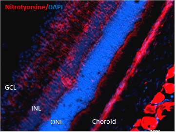
Immunohistochemistry analysis using Mouse Anti-Nitrotyrosine Monoclonal Antibody, Clone 39B6 (SMC-154). Tissue: Retinal Injury Model. Species: Mouse. Primary Antibody: Mouse Anti-Nitrotyrosine Monoclonal Antibody (SMC-154) at 1:1000. Secondary Antibody: Alexa Fluor 594 Goat Anti-Mouse (red). Courtesy of: Dr. Rajashekhar Gangaraju, University of Indiana, Department of Ophthalmology, Eugene and Marilyn Glick Eye Institute.
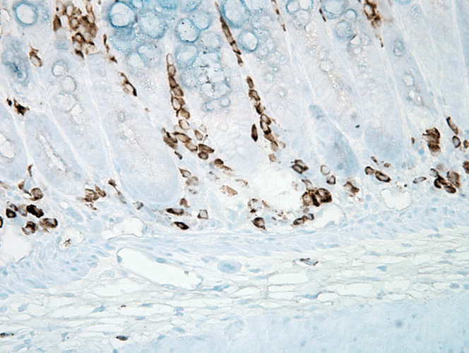
Immunohistochemistry analysis using Mouse Anti-Nitrotyrosine Monoclonal Antibody, Clone 39B6 (SMC-154). Tissue: inflamed colon. Species: Mouse. Fixation: Formalin. Primary Antibody: Mouse Anti-Nitrotyrosine Monoclonal Antibody (SMC-154) at 1:1000000 for 12 hours at 4°C. Secondary Antibody: Biotin Goat Anti-Mouse at 1:2000 for 1 hour at RT. Counterstain: Mayer Hematoxylin (purple/blue) nuclear stain at 200 µl for 2 minutes at RT. Magnification: 40x. With anti-microbial. This image was produced using an amplifying IHC wash buffer. The antibody has therefore been diluted more than is recommended for other applications.
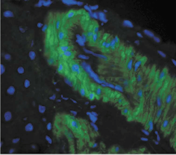
Immunohistochemistry analysis using Mouse Anti-Nitrotyrosine Monoclonal Antibody, Clone 39B6 (SMC-154). Tissue: liver tissue . Species: Rat. Primary Antibody: Mouse Anti-Nitrotyrosine Monoclonal Antibody (SMC-154) at 1:1000. Secondary Antibody: FITC Goat Anti-Mouse (green).
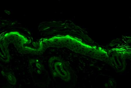
Immunohistochemistry analysis using Mouse Anti-Nitrotyrosine Monoclonal Antibody, Clone 39B6 (SMC-154). Tissue: backskin. Species: Mouse. Fixation: Bouin’s Fixative and paraffin-embedded. Primary Antibody: Mouse Anti-Nitrotyrosine Monoclonal Antibody (SMC-154) at 1:100 for 1 hour at RT. Secondary Antibody: FITC Goat Anti-Mouse (green) at 1:50 for 1 hour at RT. Backskin obtained from transgenic mice.
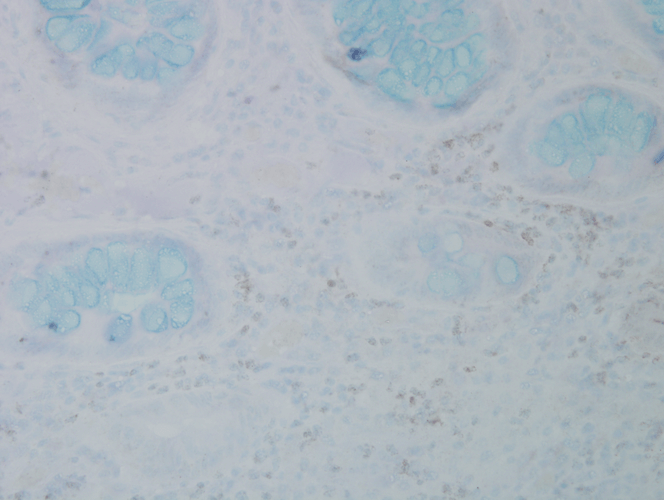
Immunohistochemistry analysis using Mouse Anti-Nitrotyrosine Monoclonal Antibody, Clone 39B6 (SMC-154). Tissue: colon carcinoma. Species: Human. Fixation: Formalin. Primary Antibody: Mouse Anti-Nitrotyrosine Monoclonal Antibody (SMC-154) at 1:25000 for 12 hours at 4°C. Secondary Antibody: Biotin Goat Anti-Mouse at 1:2000 for 1 hour at RT. Counterstain: Mayer Hematoxylin (purple/blue) nuclear stain at 200 µl for 2 minutes at RT. Magnification: 40x.
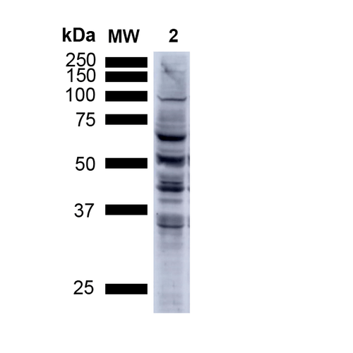
Western Blot analysis of Human A549 cells showing detection of Multiple Bands Nitrotyrosine protein using Mouse Anti-Nitrotyrosine Monoclonal Antibody, Clone 39B6 (SMC-154). Lane 1: MW ladder. Lane 2: Human A549 Cells 15 ug). Load: 15 ug. Block: 5% Skim Milk Powder in TBST. Primary Antibody: Mouse Anti-Nitrotyrosine Monoclonal Antibody (SMC-154) at 1:1000 for 2.5 hours at RT with shaking . Secondary Antibody: Goat anti-mouse IgG:HRP at 1:1000 for 1 hour at RT with shaking . Color Development: Chemiluminescent for HRP (Moss) for 5 min in RT. Predicted/Observed Size: Multiple Bands.
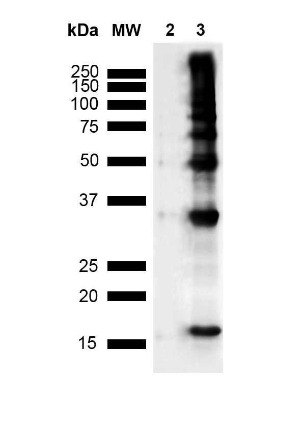
Western Blot analysis of Human Recombinant Protein showing detection of Multiple Bands Nitrotyrosine protein using Mouse Anti-Nitrotyrosine Monoclonal Antibody, Clone 39B6 (SMC-154). Lane 1: MW Ladder. Lane 2: hASYN Monomer (3.84 ug). Lane 3: Nitrosylated hASYN (3.84 ug).. Block: 5% Skim Milk Powder in TBST. Primary Antibody: Mouse Anti-Nitrotyrosine Monoclonal Antibody (SMC-154) at 1:1000 for 2 hours at RT with shaking . Secondary Antibody: Goat anti-mouse IgG:HRP at 1:4000 for 2 hour at RT with shaking . Color Development: Chemiluminescent for HRP (Moss) for 5 min in RT. Predicted/Observed Size: Multiple Bands.
 Powered by Bioz
StressMarq Biosciences :
Based on validation through cited publications.