Discovery through Partnership | Excellence through Quality


Properties
| Storage Buffer | PBS, 50% glycerol, 0.09% sodium azide *Storage buffer may change when conjugated |
| Storage Temperature | -20ºC, Conjugated antibodies should be stored according to the product label |
| Shipping Temperature | Blue Ice or 4ºC |
| Purification | Peptide Affinity Purified |
| Clonality | Polyclonal |
| Specificity | Detects ~100 kDa. |
| Cite This Product | StressMarq Biosciences Cat# SPC-640, RRID: AB_2705267 |
| Certificate of Analysis | A 1:1000 dilution of SPC-640 was sufficient for detection of ULK1 in 15 µg of Human HeLa Cell Lysates by ECL immunoblot analysis using goat anti-rabbit IgG:HRP as the secondary antibody. |
Biological Description
| Alternative Names | Unc-51 like kinase 1 (C. elegans) Antibody, Autophagy related protein 1 homolog Antibody, Autophagy-related protein 1 homolog Antibody, FLJ38455 Antibody, Serine/threonine-protein kinase ULK1 Antibody, Unc51.1 Antibody, ATG1A Antibody, UNC51, C. elegans, homolog of Antibody, EC:2.7.11.1 Antibody, Serine/threonine protein kinase Unc51.1 Antibody, FLJ46475 Antibody, ATG 1 Antibody, hATG1 Antibody, ATG1 Antibody, Unc-51-like kinase 1 Antibody, Serine/threonine protein kinase ULK1 Antibody, ULK1 Antibody, Unc 51 like kinase 1 Antibody, KIAA0722 Antibody, UNC 51 Antibody, ULK 1 Antibody, ULK1_HUMAN Antibody, UNC51 Antibody, Unc 51 (C. elegans) like kinase 1 Antibody, ATG1 autophagy related 1 homolog Antibody |
| Research Areas | Cardiovascular System, Cell Signaling, Heart, Metabolism, Mitochondrial Metabolism, Neuroscience, Protein Phosphorylation, Serine / Threonine Kinases |
| Cellular Localization | Cytoplasm, Cytosol, Preautophagosomal structure |
| Accession Number | NP_003556.1 |
| Gene ID | 8408 |
| Swiss Prot | O75385 |
| Scientific Background | UNC-51 like kinase 1 (ULK1) is widely expresed and contains an amino-terminal kinase domain followed by a central proline-serine rich domain and a highly conserved carboxy-terminal domain. It has been linked to axon growth and is essential for autophagy. Structurally, ULK1 is similar to ATG1, and it appears that both Atg1/ULK1 can bind to several ATG proteins regulating phosphorylation states and protein trafficking. |
| References |
1. Okazaki N., et al. (2000) Brain Res Mol Brain Res. 85: 1-12. 2. Young A.R., et al. (2006) J Cell Sci. 119: 3888-900. 3. Kamada Y., et al. (2000) J Cell Biol. 150: 1507-13. 4. Lee S.B, et al. (2007) EMBO Rep. 8: 360-5. 5. Hara T., et al. (2008) J Cell Biol. 181: 497-510. |
Product Images
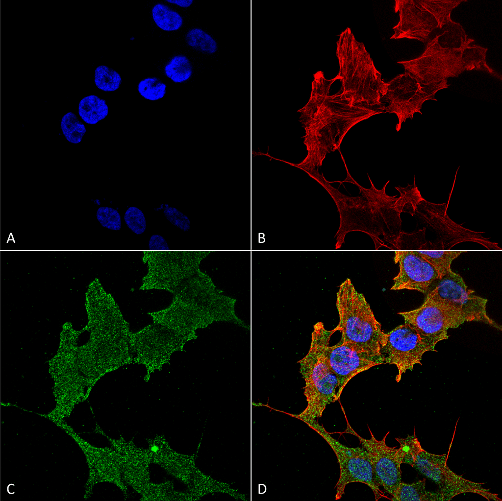
Immunocytochemistry/Immunofluorescence analysis using Rabbit Anti-ULK1 Polyclonal Antibody (SPC-640). Tissue: Neuroblastoma cell line (SK-N-BE). Species: Human. Fixation: 4% Formaldehyde for 15 min at RT. Primary Antibody: Rabbit Anti-ULK1 Polyclonal Antibody (SPC-640) at 1:100 for 60 min at RT. Secondary Antibody: Goat Anti-Rabbit ATTO 488 at 1:200 for 60 min at RT. Counterstain: Phalloidin Texas Red F-Actin stain; DAPI (blue) nuclear stain at 1:1000, 1:5000 for 60 min at RT, 5 min at RT. Localization: Cytoplasm, Preautophagosomal Structure. Magnification: 60X. (A) DAPI (blue) nuclear stain (B) Phalloidin Texas Red F-Actin stain (C) ULK1 Antibody (D) Composite.
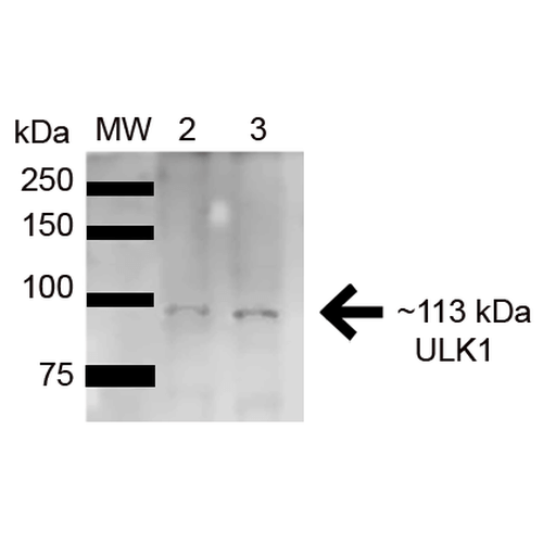
Western blot analysis of Human HeLa and HEK293Trap cell lysates showing detection of 112.6 kDa ULK1 protein using Rabbit Anti-ULK1 Polyclonal Antibody (SPC-640). Lane 1: Molecular Weight Ladder (MW). Lane 2: HeLa (15ug) . Lane 3: Human 293Trap (15ug) . Load: 15 µg. Block: 5% Skim Milk in 1X TBST. Primary Antibody: Rabbit Anti-ULK1 Polyclonal Antibody (SPC-640) at 1:1000 for 1 hour at RT. Secondary Antibody: Goat Anti-Rabbit HRP at 1:2000 for 60 min at RT. Color Development: ECL solution for 6 min in RT. Predicted/Observed Size: 112.6 kDa.
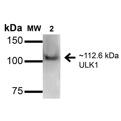
Western blot analysis of Rat Brain cell lysates showing detection of ~112.6kDa ULK1 protein using Rabbit Anti-ULK1 Polyclonal Antibody (SPC-640). Lane 1: Molecular Weight Ladder (MW). Lane 2: Rat Brain cell lysates. Load: 15 µg. Block: 2% GE Healthcare Blocker (RT, 60 minutes). Primary Antibody: Rabbit Anti-ULK1 Polyclonal Antibody (SPC-640) at 1:1000 for 16 hours at 4°C. Secondary Antibody: Goat Anti-Rabbit IgG: HRP at 1/2000 for 60 min at RT. Color Development: ECL solution for 6 min at RT. Predicted/Observed Size: ~112.6kDa.
 Powered by Bioz
Reviews
There are no reviews yet.