Discovery through Partnership | Excellence through Quality
| Product Name | HSC70 ELISA kit |
| Description |
Colorimetric detection of HSC70 |
| Species Reactivity | Avian (Zebra finch), Human |
| Platform | Microplate |
| Sample Types | Cell lysates, Serum, Tissue, Whole Blood |
| Detection Method | Colorimetric Assay |
| Assay Type | Sandwich ELISA (Enzyme-linked Immunosorbent Assay) |
| Utility | ELISA kit used to quantitate HSC70 concentration in samples. |
| Sensitivity | 1.54 ng/ml |
| Assay Range | 2.34 - 150 ng/mL |
| Incubation Time | 30 minutes |
| Number of Samples | 40 samples in duplicate |
| Other Resources | Kit Booklet Lot No. SH779113 , Kit Booklet Lot No. SH111999 , Kit Booklet Lot No. SH988614 , Kit Booklet Lot No. 131007 , Kit Booklet Lot No. 1008 , MSDS |
| Field of Use | Not for use in humans. Not for use in diagnostics or therapeutics. For in vitro research use only. |
Properties
| Storage Temperature | 4ºC and -20ºC | |||||||||||||||||||||||||||||||||||||||
| Shipping Temperature | Blue Ice | |||||||||||||||||||||||||||||||||||||||
| Product Type | ELISA Kits | |||||||||||||||||||||||||||||||||||||||
| Assay Overview | 1. Prepare Standard and samples in Standard and Sample Diluent. 2. Add 100 µL of Standard or sample to appropriate wells. 3. Cover plate with Plate Sealer and incubate at room temperature (20-25°C) for 1 hour. 4. Wash plate four times with 1X Wash Buffer. 5. Add 100 µL of Biotinylated Antibody Working Solution to each well. 6. Cover plate with Plate Sealer and incubate at room temperature for 1 hour. 7. Wash plate four times with 1X Wash Buffer. 8. Add 100 µL of Streptavidin-HRP Working Solution to each well. 9. Cover plate with Plate Sealer and incubate at room temperature for 30 minutes. 10. Wash plate four times with 1X Wash Buffer. 11. Add 100 µL of TMB Substrate to each well. 12. Develop the plate in the dark at room temperature for 30 minutes. 13. Stop reaction by adding 100 µL of Stop Solution to each well. 14. Measure absorbance on a plate reader at 450 nm. | |||||||||||||||||||||||||||||||||||||||
| Kit Overview |
|
|||||||||||||||||||||||||||||||||||||||
| Cite This Product | HSC70 ELISA Kit (StressMarq Biosciences Inc., Victoria BC CANADA, Catalog # SKT-106) |
Biological Description
| Alternative Names | HSC54 ELISA Kit, HSC71 ELISA Kit, HSC73 ELISA Kit, HSP71 ELISA Kit, HSP73 ELISA Kit, HSPA10 ELISA Kit, HSPA8 ELISA Kit, LAP1 ELISA Kit, NIP71 ELISA Kit |
| Research Areas | Cancer, Heat Shock |
| Scientific Background | HSP70 genes encode abundant heat-inducible 70-kDa HSPs (HSP70s). In most eukaryotes HSP70 genes exist as part of a multigene family. They are found in most cellular compartments of eukaryotes including nuclei, mitochondria, chloroplasts, the endoplasmic reticulum and the cytosol, as well as in bacteria. The genes show a high degree of conservation, having at least 5O% identity (2). The N-terminal two thirds of HSP70s are more conserved than the C-terminal third. HSP70 binds ATP with high affinity and possesses a weak ATPase activity which can be stimulated by binding to unfolded proteins and synthetic peptides (3). When HSC70 (constitutively expressed) present in mammalian cells was truncated, ATP binding activity was found to reside in an N-terminal fragment of 44 kDa which lacked peptide binding capacity. Polypeptide binding ability therefore resided within the C-terminal half (4). The structure of this ATP binding domain displays multiple features of nucleotide binding proteins (5). When cells are subjected to metabolic stress (e.g., heat shock) a member of the HSP 70 family, HSP 70 (HSP72), is expressed; HSP 70 is highly related to HSC70 (>90% sequence identity). Constitutively expressed HSC70 rapidly forms a stable complex with the highly inducible HSP70 in cells following heat shock. The interaction of HSC70 with HSP 70 is regulated by ATP. These two heat shock proteins move together in the cell experiencing stress. Furthermore, research on HSC70 has implicates it with a role in facilitating the recovery of centrosomal structure and function after heat shock (6). |
| References |
1. Brown C.L., et al. (1993) J.Cell Biol., 120 (5): 1101-1112. 2. Boorstein W.R., Ziegelhoffer T., and Craig E.A. (1993) J. Mol. Evol. 38(1): 1-17. 3. Rothman J. (1989) Cell 59: 591-601. 4. DeLuca-Flaherty, et al. (1990) Cell 62: 875-887. 5. Bork P., Sander C., and Valencia A. (1992) Proc. Natl Acad. Sci. USA 89: 7290-7294. 6. Brown C.L., et al. (1996) J. Biol. Chem. 271(2): 833-840. |
Product Images
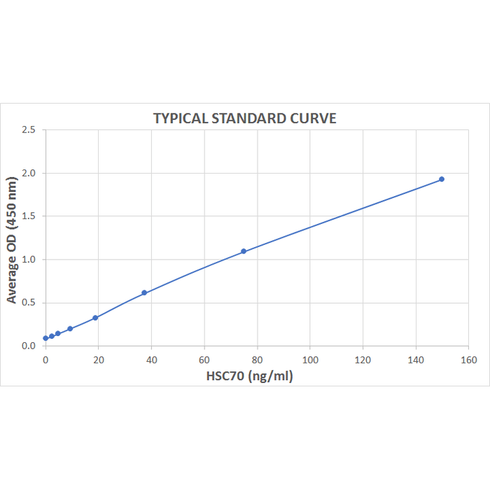
Typical Standard Curve for the HSC70 ELISA kit (Enzyme-Linked Immunosorbent Assay) StressXpress® – SKT-106. Assay Type: Sandwich ELISA. Detection Method: Colorimetric Assay. Assay Range: 2.34 – 150 ng/mL.
 Powered by Bioz
StressMarq Biosciences :
Based on validation through cited publications.