Discovery through Partnership | Excellence through Quality


Properties
| Storage Buffer | PBS pH7.4, 50% glycerol, 0.09% sodium azide *Storage buffer may change when conjugated |
| Storage Temperature | -20ºC, Conjugated antibodies should be stored according to the product label |
| Shipping Temperature | Blue Ice or 4ºC |
| Purification | Protein G Purified |
| Clonality | Monoclonal |
| Clone Number | 5D12-A12 |
| Isotype | IgG2b Kappa |
| Specificity | Detects ~27kDa. Has no cross-reactivity to Alpha B crystallin. Very limited cross-reactivity to other species. |
| Cite This Product | StressMarq Biosciences Cat# SMC-161, RRID: AB_2248586 |
| Certificate of Analysis | 0.5 µg/ml of SMC-161 was sufficient for detection of HSP27 in 10 µg of HeLa lysate by colorimetric immunoblot analysis using Goat anti-mouse IgG:HRP as the secondary antibody. |
Biological Description
| Alternative Names | 28kDa heat shock protein Antibody, CMT2F Antibody, HSP25 Antibody, HSP27 Antibody, HSP28 Antibody, HSPB1 Antibody, SRP27 Antibody |
| Research Areas | Actin, Actin Assembly, Atherosclerosis, Cancer, Cardiovascular System, Cell Signaling, Chaperone Proteins, Contractility, Cytoskeleton, Heart, Heat Shock, Microfilaments, Microtubules, Protein Trafficking, Tumor Biomarkers |
| Cellular Localization | Cytoplasm, Cytoskeleton, Nucleus, Spindle |
| Accession Number | NP_001531.1 |
| Gene ID | 3315 |
| Swiss Prot | P04792 |
| Scientific Background | HSP27s belong to an abundant and ubiquitous family of small heat shock proteins (sHSP). It is an important HSP found in both normal human cells and cancer cells. The basic structure of most sHSPs is a homologous and highly conserved amino acid sequence, with an α-crystallin domain at the C-terminus and the WD/EPF domain at the less conserved N-terminus. This N-terminus is essential for the development of high molecular oligomers (1, 2). HSP27-oligomers consist of stable dimers formed by as many as 8-40 HSP27 protein monomers (3). The oligomerization status is connected with the chaperone activity: aggregates of large oligomers have high chaperone activity, whereas dimers have no chaperone activity (4). HSP27 is localized to the cytoplasm of unstressed cells but can redistribute to the nucleus in response to stress, where it may function to stabilize DNA and/or the nuclear membrane. Other functions include chaperone activity (as mentioned above), thermo tolerance in vivo, inhibition of apoptosis, and signal transduction. Specifically, in vitro, it acts as an ATP-independent chaperone by inhibiting protein aggregation and by stabilizing partially denatured proteins, which ensures refolding of the HSP70 complex. HSP27 is also involved in the apoptotic signaling pathway because it interferes with the activation of cytochrome c/Apaf-1/dATP complex, thereby inhibiting the activation of procaspase-9. It is also hypothesized that HSP27 may serve some role in cross-bridge formation between actin and myosin (5). And finally, HSP27 is also thought to be involved in the process of cell differentiation. The up-regulation of HSP27 correlates with the rate of phosphorylation and with an increase of large oligomers. It is possible that HSP27 may play a crucial role in termination of growth (6). For more information visit our HSP27 Scientific Resource Guide at http://www.HSP27.com. |
| References |
1. Kim K.K., Kim R., and Kim, S. (1998) Nature 394(6693): 595-599. 2. Van Montfort R., Slingsby C., and Vierling E. (2001) Addv Protein Chem. 59: 105-56. 3. Ehrnsperger M., Graber S., Gaestel M. and Buchner J. (1997) EMBO J. 16: 221-229. 4. Ciocca D.R., Oesterreich S., Chamness G.C., McGuire W.L., and Fugua S.A. (1993) J Natl Cancer Inst. 85 (19): 1558-70. 5. Sarto C., Binnz P.A., and Mocarelli P. (2000) Electrophoresis. 21(6): 1218-26. 6. Arrigo A.P. (2005) J Cell Biochem. 94(2): 241-6. |
Product Images
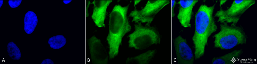
Immunocytochemistry/Immunofluorescence analysis using Mouse Anti-Hsp27 Monoclonal Antibody, Clone 5D12-A3 (SMC-161). Tissue: Heat Shocked cervical cancer cells (HeLa). Species: Human. Fixation: 2% Formaldehyde for 20 min at RT. Primary Antibody: Mouse Anti-Hsp27 Monoclonal Antibody (SMC-161) at 1:100 for 12 hours at 4°C. Secondary Antibody: FITC Goat Anti-Mouse (green) at 1:200 for 2 hours at RT. Counterstain: DAPI (blue) nuclear stain at 1:40000 for 2 hours at RT. Localization: Cytoplasm. Nucleus. Magnification: 100x. (A) DAPI (blue) nuclear stain. (B) Anti-Hsp27 Antibody. (C) Composite. Heat Shocked at 42°C for 1h.
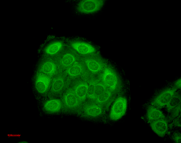
Immunocytochemistry/Immunofluorescence analysis using Mouse Anti-Hsp27 Monoclonal Antibody, Clone 5D12-A3 (SMC-161). Tissue: HaCaT cells. Species: Human. Fixation: Cold 100% methanol for 10 minutes at -20°C. Primary Antibody: Mouse Anti-Hsp27 Monoclonal Antibody (SMC-161) at 1:100 for 1 hour at RT. Secondary Antibody: FITC Goat Anti-Mouse (green) at 1:50 for 1 hour at RT. Localization: Dull heterogeneous staining, some perinuclear, some nuclear and some cytoplasmic staining .
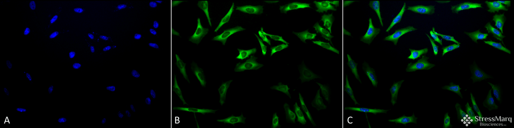
Immunocytochemistry/Immunofluorescence analysis using Mouse Anti-Hsp27 Monoclonal Antibody, Clone 5D12-A3 (SMC-161). Tissue: Heat Shocked cervical cancer cells (HeLa). Species: Human. Fixation: 2% Formaldehyde for 20 min at RT. Primary Antibody: Mouse Anti-Hsp27 Monoclonal Antibody (SMC-161) at 1:100 for 12 hours at 4°C. Secondary Antibody: FITC Goat Anti-Mouse (green) at 1:200 for 2 hours at RT. Counterstain: DAPI (blue) nuclear stain at 1:40000 for 2 hours at RT. Localization: Cytoplasm. Nucleus. Magnification: 20x. (A) DAPI (blue) nuclear stain. (B) Anti-Hsp27 Antibody. (C) Composite. Heat Shocked at 42°C for 1h.
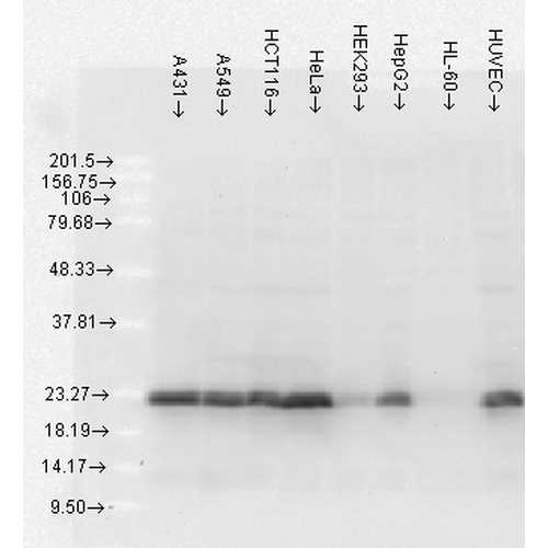
Western Blot analysis of Human Cell lysates showing detection of Hsp27 protein using Mouse Anti-Hsp27 Monoclonal Antibody, Clone 5D12-A3 (SMC-161). Load: 15 µg. Block: 1.5% BSA for 30 minutes at RT. Primary Antibody: Mouse Anti-Hsp27 Monoclonal Antibody (SMC-161) at 1:1000 for 2 hours at RT. Secondary Antibody: Sheep Anti-Mouse IgG: HRP for 1 hour at RT.
 Powered by Bioz
Evgeny Mymrikov :
Read the full review at pAbmAbs.com
StressMarq Biosciences :
Based on validation through cited publications.