HSP70 Resource Guide
HSP70 Resources
 HSP70 StressXpress® ELISA Kit |
 HSP70 High-Sensitivity StressXpress® ELISA Kit |
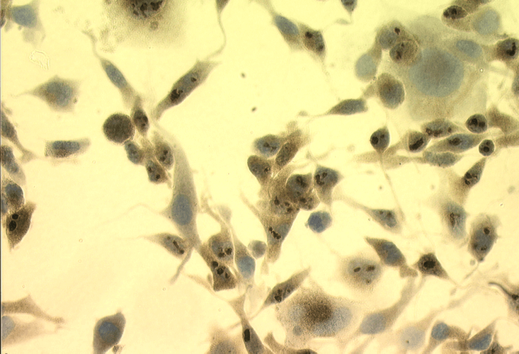
Video: Cell Membrane HSP70 Antibody for Cancer Research
StressMarq’s HSP70 antibody, clone 1H11 (catalog# SMC-249) is unique from other commercially available Hsp70 antibodies. It can bind to the extracellular region of cell membrane embedded HSP70 protein, allowing researchers to differentiate between intracellular and cell membrane embedded HSP70 across cancer cell types.
Product and Research Area Links
Heat Shock Proteins (HSPs) were initially discovered as proteins that were markedly induced by heat shock and other chemical or physical stresses. Since that time they have viewed as molecular chaperones, guiding and modifying the structures of proteins by their intrinsic ability to re-fold them from a denatured to a native structure. These interactions and the balance toward fully-natured functional proteins necessitates that the HSPs be abundant. As a result, the HSP27, HSP40, HSP70 and HSP110 genes have evolved to be particularly efficient for mass synthesis after or during stress with powerful transcriptional activation, efficient messenger RNA (mRNA) stabilization, and selective mRNA translation. HSP27, HSP70, HSP90, and HSP110 increase to become the dominantly expressed proteins after stress. The types of stress are numerous, but include heat shock, metal toxicity, nutrient deprivation, oxidative stress as well as numerous disease states of which cancer has received the most attention.
HSPs are regulated by transcription factors that belong to the heat shock factor family, which allows rapid transcriptional activation as well as ensuring a prompt shut-off post recovery. These heat shock factors include HSF1, HSF2 and HSF4. However, a significant amount of HSPs are constitutively expressed. Both induced or constitutively expressed HSPs function is several ways, and have the ability to form complexes with client proteins. These complexes are themselves directly involved in protein folding in the cytosol, endoplasmic reticulum (ER) and mitochondria, the intracellular transport of proteins, protein repair and degradation (of environmentally damaged proteins), regulatory protein control, and the re-folding of misfolded proteins. Examples of such complexes include HSP10 and HSP60 complexes that mediate protein folding and HSP70- and HSP90-containing complexes that are involved in both generic protein-folding pathways and in specific association with key regulatory proteins within the cell. HSP90 plays a particularly versatile role in cell regulation – it is the ability to form complexes with a large number of cellular kinases, transcription factors, and other molecules that has led to the protein becoming a drug target of considerable interest. The HSPs have been generally classified into several classes depending on their molecular weights, see Table 1*.
Table 1 (HSP Families)*
| Protein Name | Localization | Function |
|---|---|---|
| HSP104 | Cytoplasm | Releases proteins from aggregates |
| HSP90α and HSP90ß | Cytoplasm | Prevents protein aggregation, enables protein stabilization and trafficking, facilitates activation of numerous regulated proteins |
| Grp94 | Endoplasmic reticulum | Quality control of protein processing in the endoplasmic reticulum |
| TRAP / HSP75 | Mitochondria | Unclear |
| HSP70 / Hsc70 | Cytoplasm | Prevents protein aggregation, aids protein folding |
| Grp78 / Bip | Endoplasmic reticulum | Protein import and folding in the endoplasmic reticulum |
| HSP60 / Chaperonins | Cytoplasm and mitochondria | Prevents protein aggregation, aids protein folding |
| HSP47 | Endoplasmic reticulum | Facilitates the folding and assembly of pro-collagen molecules, retaining unfolded molecules within the ER, and assisting the transport of correctly folded molecules from the ER to the Golgi apparatus. |
| HSP40 / HDJ2 | Cytoplasm | Helps protein folding as a co-chaperone of HSP70 |
| HSP32 (HO-1; small HSPs) | Endoplasmic reticulum, plasma membrane and mitochondria | Catalyzes first step of heme degradation to bilirubin, which has antioxidant properties. |
| HSP27 / HSP25 (small HSPs) | Endoplasmic reticulum | Prevents protein aggregation, may have role in cell growth and differentiation |
| Alpha B crystallin (small HSPs) | Cytoplasm | Major eye lens protein. It inhibits TRAIL induced apoptosis in cancer. It confers a cytoprotective effect by suppressing aggregation of denatured proteins. It is constitutively expressed, often at high levels, in human cancers, including gliomas, breast, prostate and renal cell carcinomas. |
* Adapted from Annals in Oncology, 14 1169-1176, 2003
HSP70 (also called heat shock protein 70 or HSP72) has a wide range of diverse functions. It is inducibly expressed, whereas Hsc70 (heat shock cognate 70) – sharing a very high degree of homology and function – is not. Hsc70 (also called HSP73) is constitutively expressed. Generally HSP70 it is seen as a mostly an anti-apoptotic protein. It interacts with the intrinsic and extrinsic pathways of apoptosis at several junctions and inhibits cell death through chaperone dependent as well as independent activities. HSP70 protects cells from cytotoxicity induced by TNF, monocytes, oxidative stress, chemotherapeutic agents, ceramide and radiation. The apoptotic events generated by nitric oxide and heat stress triggers translocation of Bax from cytoplasm to the mitochondria, which is inhibited by over-expression of HSP70. Downstream in the pathway, HSP70 also inhibits formation of a functional apoptosome complex by direct interaction with Apaf-1. It prevents late caspase dependent events such as activation of cytosolic phospholipase A2 and changes in nuclear morphology; it can also protect cells from forced expression of caspase-3. HSP70 can, independent of its chaperoning activity, inhibit JNK mediated cell death, by suppressing JNK phosphorylation either directly and/or through the upstream SEK kinase.
HSP70 and HSP90 have been identified to have a peptide carrier function. They have been found to be involved in the cross-presentation of tumor-derived peptides on MHC class I molecules. More diverse functions have been reported, however – the binding of mycobacterial HSP70 to CD40 was found to mediate a calcium-dependent cell signaling and the release of CC chemokines, pro-inflammatory cytokines, and nitric oxide, whereas mammalian HSP70s were found to facilitate receptor-mediated endocytosis.
HSP70 and HSP90 are able signal danger to the cell even in the absence of immunogenic peptides. Tumor cells themselves have been identified as a source of extracellular HSP70s, and after treatment with IFN-Hsc70 has been observed. Furthermore, pro-inflammatory cytokines are released after the interactions of peptide-free HSP70 with CD14 and TLR2/4 on antigen-presenting cells. The process is initiated by the translocation of NF-B into the nucleus. The cytokine release triggers the stimulation of innate immune system.
HSP70 also competes with the CD40 ligand for binding to antigen-presenting cells. In addition HSP70s appear to be involved in the stimulation of the migration of dendritic cells to the draining lymph nodes and in maturation of those cells They show up-regulation of CD86, CD83, and CD40 after contact with HSP70 MHC class II. However, the role of HSPs as cytokine-like proteins appears to be due, at least in part to contaminating levels of LPS or bacterial lipoprotein contamination in the HSPs preparations. It is also clear that many HSPs can bind LPS. Reduction of LPS levels reduce the stimulatory capacity of HSP70s and HSP90s toward dendritic cells.
Natural Killer (NK) cells are important effector cells of the innate immune system. HSP70 was identified as being a trigger factor for NK cells with a high CD94 surface density. After mapping the interaction if was found that only a 14-mer peptide – T-K-D-N-N-L-L-G-R-F-E-L-S-G (TKD; AA450-463) from the C-terminal domain was required for immuno-stimulus. When incubated with cytokines and HSP70 or the TKD peptide the cell surface density of NK receptors increases, including CD94. Further blocking studies showed the importance of CD94 in the interaction of NK cells with HSP70 on tumor cells. Screening of human tumor biopsies have revealed that HSP70 is often present of plasma membranes of the colon, lungs, pancreas, head and neck and metastases thereof. Typical or normal tissue samples are found to be HSP70-negative. Interestingly, the cell surface densities (from tumor tissues) of HSP70 can be further increased by the administration of reagents (such as membrane-interactive alkyl-lysophospholipids, cytostatic drugs, including taxoides and vincristine sulfate, cyclooxygenase (COX-1/2) inhibitors, acetylsalicylic acid, insulin sensitizers, or subjecting the samples to hyperthermia, radiation, and photodynamic therapy. This heightened HSP70 density correlates with an increased sensitivity to NK cell -mediated cell death, suggesting an NK cell based therapeutic capacity further increased by stimulation with chemical or chemical means.
It has been shown that HSP70 induction is important in neuronal survival after a stroke, which correlates with the classical observations of the importance of HSPs and their role cardio-protection. HSP70 is also able to improve tissue transplantation efficiency, and eases the serious implications of chronic diseases such as diabetes (resulting form studies using HSP70 inducers such as bimoclomol and BRX-220). Other neurological diseases including Huntington’s, Parkinson’s and Alzheimer’s or neurological trauma cases show beneficial effects if over-expression of HSP70 (and HSP40) are present. Inducers of HSP70 are numerous, and include stannous chloride (improving the success rate of tissue transplantations), geranyl-geranyl acetone (protects neurons against cerebral ischemia), the antiulcer drug carbenoxolone and probably the best known one – aspirin, which enhances HSP70 synthesis.