Discovery through Partnership | Excellence through Quality


Properties
| Storage Buffer | PBS pH7.4, 50% glycerol, 0.1% sodium azide *Storage buffer may change when conjugated |
| Storage Temperature | -20ºC, Conjugated antibodies should be stored according to the product label |
| Shipping Temperature | Blue Ice or 4ºC |
| Purification | Protein G Purified |
| Clonality | Monoclonal |
| Clone Number | C92F3A-5 |
| Isotype | IgG1 |
| Specificity | Detects ~70kDa. Does not cross-react with HSC70 (HSP73). |
| Cite This Product | HSP70 Antibody (StressMarq Biosciences | Victoria, BC CANADA, Catalog# SMC-100, RRID: AB_854199) |
| Certificate of Analysis | 1 µg/ml of SMC-100 was sufficient for detection of HSP70 in 20 µg of heat shocked HeLa cell lysate by colorimetric immunoblot analysis using Goat anti-mouse IgG:HRP as the secondary antibody. |
Biological Description
| Alternative Names | HSPA1A, HSPA1B, HSPA1, HSP70, HSP70-1, HSP70.1, HSP70-2, HSP72, HSP73, HSX70, Heat shock 70 kDa protein 1A, Heat shock 70 kDa protein 1B |
| Research Areas | Cancer, Cell Signaling, Chaperone Proteins, Heat Shock, Protein Trafficking, Tumor Biomarkers |
| Cellular Localization | Cytoplasm |
| Accession Number | NP_005336.3 |
| Gene ID | 3303 |
| Swiss Prot | P0DMV8/P0DMV9 |
| Scientific Background |
HSP70 proteins are a highly conserved family of 70-kDa molecular chaperones encoded by a multigene family in most eukaryotes and prokaryotes. Found in nearly all cellular compartments—including the cytosol, nucleus, mitochondria, endoplasmic reticulum, and chloroplasts—HSP70s are constitutively expressed and strongly upregulated in response to cellular stress. These chaperones play a vital role in protein homeostasis by binding to nascent polypeptides and partially folded or misfolded proteins, preventing aggregation and facilitating proper folding. HSP70s exhibit high-affinity ATP binding and weak ATPase activity, which is stimulated upon interaction with unfolded substrates. ATP hydrolysis triggers conformational changes that regulate substrate binding and release, enabling dynamic cycles of protein folding and refolding. Structurally, the N-terminal domain of HSP70 is responsible for ATP binding, while the C-terminal domain mediates substrate interaction. This modular design allows HSP70s to coordinate with co-chaperones such as HSP40, HIP, HOP, and BAG-1, integrating folding with degradation and transport pathways. In neurodegenerative disease research, HSP70 is of particular interest due to its ability to counteract protein misfolding and aggregation—hallmarks of disorders like Alzheimer’s, Parkinson’s, and Huntington’s disease. By stabilizing toxic intermediates and promoting their clearance, HSP70 enhances neuronal survival and resilience under proteotoxic stress. |
| References |
1. Welch W.J. and Suhan J.P. (1986) J Cell Biol. 103: 2035-2050. 2. Boorstein W. R., Ziegelhoffer T. & Craig E. A. (1993) J.Mol. Evol. 38(1): 1-17. 3. Rothman J. (1989) Cell 59: 591-601. 4. DeLuca-Flaherty et al. (1990) Cell 62: 875-887. 5. Bork P., Sander C. & Valencia A. (1992) Proc. Nut1 Acad. Sci. USA 89: 7290-7294. 6. Fink A.L. (1999) Physiol. Rev. 79: 425-449. 7. Galan A., et al. (2000) J. Biol. Chem. 275: 11418-11424. 8. Kondo T., et al. (2000) J. Biol. Chem. 275: 8872-8879. 9. Misaki T., et al. (1994) Clin. Exp. Immun. 98: 234-239. 10. Pockley A.G., et al. (1998) Immunol. Invest. 27: 367-377. 11. Moon I.S., et al. (2001) Cereb Cortex 11(3): 238-248. 12. Dressel et al. (2000) J. Immunol. 164: 2362-2371. 13. Verma A.K., et al. (2007) Fish and Shellfish Immunology. 22(5): 547-555. 14. Banduseela V.C., et al. (2009) Physiol Genomics. 39(3): 141-159. |
Product Images
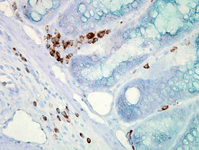
Immunohistochemistry analysis using Mouse Anti-Hsp70 Monoclonal Antibody, Clone C92F3A-5 (SMC-100). Tissue: colon carcinoma. Species: Mouse. Fixation: Formalin. Primary Antibody: Mouse Anti-Hsp70 Monoclonal Antibody (SMC-100) at 1:10000 for 12 hours at 4°C. Secondary Antibody: Biotin Goat Anti-Mouse at 1:2000 for 1 hour at RT. Counterstain: Mayer Hematoxylin (purple/blue) nuclear stain at 200 µl for 2 minutes at RT. Localization: Inflammatory cells. Magnification: 40x. This image was produced using an amplifying IHC wash buffer. The antibody has therefore been diluted more than is recommended for other applications.
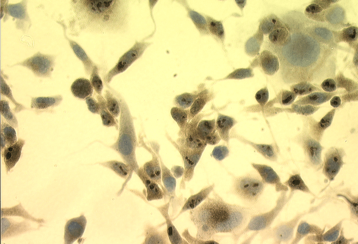
Immunocytochemistry/Immunofluorescence analysis using Mouse Anti-Hsp70 Monoclonal Antibody, Clone C92F3A-5 (SMC-100). Tissue: Heat Shocked Melanoma cells. Species: Mouse. Fixation: Formalin. Primary Antibody: Mouse Anti-Hsp70 Monoclonal Antibody (SMC-100) at 1:1000 for 16 hours at RT. Secondary Antibody: Biotin Goat Anti-Mouse. Courtesy of: Dr. Ewa Malusecka, Maria Sklodowska-Curie Memorial Cancer Centre and Inst. Of Oncology, Poland.
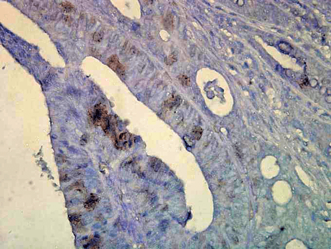
Immunohistochemistry analysis using Mouse Anti-Hsp70 Monoclonal Antibody, Clone C92F3A-5 (SMC-100). Tissue: colon carcinoma. Species: Human. Fixation: Formalin. Primary Antibody: Mouse Anti-Hsp70 Monoclonal Antibody (SMC-100) at 1:10000 for 12 hours at 4°C. Secondary Antibody: Biotin Goat Anti-Mouse at 1:2000 for 1 hour at RT. Counterstain: Mayer Hematoxylin (purple/blue) nuclear stain at 200 µl for 2 minutes at RT. Localization: Inflammatory cells. Magnification: 40x. This image was produced using an amplifying IHC wash buffer. The antibody has therefore been diluted more than is recommended for other applications.
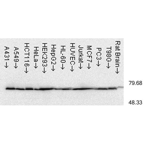
Western Blot analysis of Human cell lysates from various cell lines showing detection of Hsp70 protein using Mouse Anti-Hsp70 Monoclonal Antibody, Clone C92F3A-5 (SMC-100). Load: 15 µg. Block: 1.5% BSA for 30 minutes at RT. Primary Antibody: Mouse Anti-Hsp70 Monoclonal Antibody (SMC-100) at 1:1000 for 2 hours at RT. Secondary Antibody: Sheep Anti-Mouse IgG: HRP for 1 hour at RT.
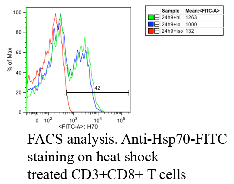
Fluorescence Activated Cell Sorting analysis using Mouse Anti-Hsp70: FITC Monoclonal Antibody, Clone C92F3A-5 (SMC-100). Tissue: Heat Shocked CD3+ CD8+ T cells . Species: Mouse. Primary Antibody: Mouse Anti-Hsp70: FITC Monoclonal Antibody (SMC-100) at 1:1000. Courtesy of: Cheryl Cameron, Vaccine and Gene Therapy Instit. Florida.
 Powered by Bioz
StressMarq Biosciences :
Based on validation through cited publications.