Advancing the Frontiers of Neurodegenerative Disease Research


Properties
| Storage Buffer | PBS, 50% glycerol, 0.09% sodium azide *Storage buffer may change when conjugated |
| Storage Temperature | -20ºC, Conjugated antibodies should be stored according to the product label |
| Shipping Temperature | Blue Ice or 4ºC |
| Purification | Protein G Purified |
| Clonality | Monoclonal |
| Clone Number | LK1 |
| Isotype | IgG1 |
| Specificity | Detects ~60kDa. |
| Cite This Product | HSP60 Antibody (StressMarq Biosciences | Victoria, BC CANADA, Catalog# SMC-110, RRID: AB_2121277) |
| Certificate of Analysis | 0.05 µg/ml of SMC-110 was sufficient for detection of HSP60 in 20 µg of heat shocked HeLa cell lysate by colorimetric immunoblot analysis using goat anti-mouse IgG as the secondary antibody. |
Biological Description
| Alternative Names | HSPD1, HSP60, 60 kDa heat shock protein, mitochondrial, Chaperonin 60, CPN60, HuCHA60, Heat shock protein family D member 1, GroEL homolog, mitochondrial, GROEL, HLD4, HSP 60, HSP65, SPG 13 |
| Research Areas | Cancer, Cell Signaling, Chaperone Proteins, Heat Shock, Organelle Markers, Protein Trafficking, Tags and Cell Markers |
| Cellular Localization | Mitochondrion, Mitochondrion Matrix |
| Accession Number | NP_002147.2 |
| Gene ID | 3329 |
| Swiss Prot | P10809 |
| Scientific Background |
HSP60, also known as Cpn60 or GroEL in prokaryotes, is a highly conserved molecular chaperone essential for protein folding and cellular homeostasis. Present in both prokaryotic and eukaryotic cells, HSP60 prevents protein misfolding and aggregation during biogenesis and under stress conditions. In mammals, HSP60 is localized to the mitochondria, where it partners with its co-chaperonin HSP10 to facilitate the proper folding and assembly of mitochondrial proteins. Structurally, HSP60 forms homo-oligomeric complexes of 7 or 14 subunits, exhibiting ATPase activity and reversible dissociation in the presence of Mg²⁺ and ATP. Its evolutionary conservation is underscored by the ability of human HSP60-HSP10 to functionally replace the bacterial GroEL-GroES system in engineered E. coli strains. Beyond its canonical role in mitochondrial proteostasis, HSP60 has been implicated in immune regulation and cellular stress responses. Elevated levels of HSP60 have been associated with several chronic diseases, including autoimmune disorders, coronary artery disease, diabetes, and neurodegenerative conditions such as Alzheimer’s disease and multiple sclerosis. In neuroscience, HSP60’s role in maintaining mitochondrial integrity is particularly significant, as mitochondrial dysfunction is a central feature of many neurodegenerative diseases. Its dual function in protein quality control and cellular protection positions HSP60 as a promising biomarker and therapeutic target in neurodegeneration research. |
| References |
1. Hartl, F.U. (1996) Nature 381: 571-579. 2. Bukau, B. and Horwich, A.L. (1998) Cell 92: 351-366. 3. Hartl, F.U. and Hayer-Hartl, M. (2002) Science 295: 1852- 1858. 4. Jindal, S., et al. (1989) Molecular and Cellular Biology 9: 2279-2283. 5. La Verda, D., et al (1999) Infect Dis. Obstet. Gynecol. 7: 64-71. 6. Itoh, H. et al. (2002) Eur. J. Biochem. 269: 5931-5938. 7. Gupta, S. and Knowlton, A.A. J. Cell Mol Med. 9: 51-58. 8. Deocaris, C.C. et al. (2006) Cell Stress Chaperones 11: 116-128. 9. Lai, H.C. et al. (2007) Am. J. Physiol. Endocrinol. Metab. 292: E292-E297. 10. Gao, Y.L., et al (1995) J. of Immunology 154: 3548-3556. 11. Neuer, A., et al (1997) European Society for Human Reproduction and Embryology 12(5):925-929. 12. Bason, C., et al (2003) Lancet 362(9400): 1971-1977. |
Product Images
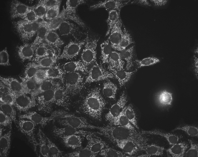
Immunocytochemistry/Immunofluorescence analysis using Mouse Anti-Hsp60 Monoclonal Antibody, Clone LK-1 (SMC-110). Tissue: HaCaT cells. Species: Human. Fixation: Cold 100% methanol at -20°C for 10 minutes. Primary Antibody: Mouse Anti-Hsp60 Monoclonal Antibody (SMC-110) at 1:100 for 1 hour at RT. Secondary Antibody: FITC Goat Anti-Mouse (green) at 1:50 for 1 hour at RT. Localization: Cytoplasmic Staining.
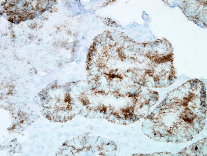
Immunohistochemistry analysis using Mouse Anti-Hsp60 Monoclonal Antibody, Clone LK-1 (SMC-110). Tissue: colon carcinoma. Species: Human. Fixation: Formalin. Primary Antibody: Mouse Anti-Hsp60 Monoclonal Antibody (SMC-110) at 1:100000 for 12 hours at 4°C. Secondary Antibody: Biotin Goat Anti-Mouse at 1:2000 for 1 hour at RT. Counterstain: Mayer Hematoxylin (purple/blue) nuclear stain at 200 µl for 2 minutes at RT. Localization: Inflammatory cells. Magnification: 40x. This image was produced using an amplifying IHC wash buffer. The antibody has therefore been diluted more than is recommended for other applications.
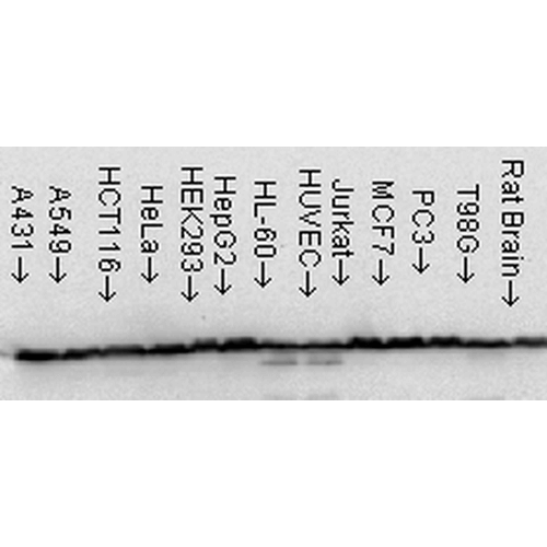
Western Blot analysis of Human Cell line lysates showing detection of Hsp60 protein using Mouse Anti-Hsp60 Monoclonal Antibody, Clone LK-1 (SMC-110). Load: 15 µg. Block: 1.5% BSA for 30 minutes at RT. Primary Antibody: Mouse Anti-Hsp60 Monoclonal Antibody (SMC-110) at 1:1000 for 2 hours at RT. Secondary Antibody: Sheep Anti-Mouse IgG: HRP for 1 hour at RT.
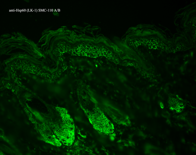
Immunohistochemistry analysis using Mouse Anti-Hsp60 Monoclonal Antibody, Clone LK-1 (SMC-110). Tissue: backskin. Species: Mouse. Fixation: Bouin’s Fixative and paraffin-embedded. Primary Antibody: Mouse Anti-Hsp60 Monoclonal Antibody (SMC-110) at 1:100 for 1 hour at RT. Secondary Antibody: FITC Goat Anti-Mouse (green) at 1:50 for 1 hour at RT. Localization: Epidermis.
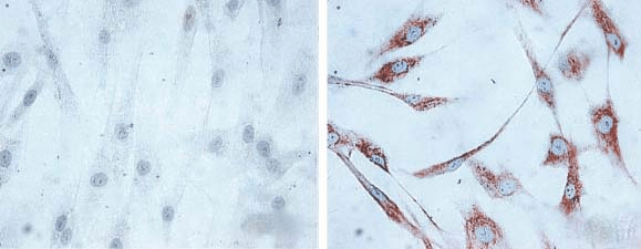
Immunocytochemistry/Immunofluorescence analysis using Mouse Anti-Hsp60 Monoclonal Antibody, Clone LK1, (SMC-110). Tissue: skin Fibroblasts. Species: Human. Fixation: Cold 100% methanol for 30 minutes at -20°C . Primary Antibody: Mouse Anti-Hsp60 Monoclonal Antibody (SMC-110) at 1:1000 for 1 hour at RT. Secondary Antibody: DAKO LSAB2 streptavidin-peroxidase system. Counterstain: Mayer Hematoxylin (purple/blue) nuclear stain. Left: control; Right: 24 hours after 7th passage of senescence. Courtesy of: Valentina di Felice, University of Palermo, Italy.
 Powered by Bioz
StressMarq Biosciences :
Based on validation through cited publications.