Discovery through Partnership | Excellence through Quality


Properties
| Storage Buffer | PBS pH7.4, 50% glycerol, 0.09% sodium azide *Storage buffer may change when conjugated |
| Storage Temperature | -20ºC, Conjugated antibodies should be stored according to the product label |
| Shipping Temperature | Blue Ice or 4ºC |
| Purification | Protein G Purified |
| Clonality | Monoclonal |
| Clone Number | 10H8 |
| Isotype | IgG1 |
| Specificity | Detects ~85kDa (unstressed cell lysates), and~95kDa (heat shocked cell lysates). |
| Cite This Product | HSF1 Antibody (StressMarq Biosciences | Victoria, BC CANADA, Catalog# SMC-118, RRID: AB_2120255) |
| Certificate of Analysis | 1 µg/ml of SMC-118 was sufficient for detection of HSF1 in 20 µg of heat shocked HeLa cell lysate by ECL immunoblot analysis using Goat anti-rat IgG: HRP as the secondary antibody |
Biological Description
| Alternative Names | HSF1, Heat shock factor protein 1, Heat shock transcription factor 1, HSTF1, HSF 1 |
| Research Areas | Cancer, Cardiovascular System, Cell Signaling, Epigenetics and Nuclear Signaling, Heart, Heat Shock |
| Cellular Localization | Cytoplasm, Nucleus |
| Accession Number | NP_032322.1 |
| Gene ID | 15499 |
| Swiss Prot | P38532 |
| Scientific Background |
Heat Shock Factor 1 (HSF1) is a key transcriptional regulator that orchestrates the cellular response to proteotoxic stress by activating genes involved in protein folding, degradation, and quality control. As the primary mediator of the heat shock response, HSF1 binds to heat shock elements (HSEs) in the promoters of heat shock protein (HSP) genes, driving the expression of molecular chaperones essential for maintaining proteostasis. Structurally, HSF1 contains a conserved N-terminal DNA-binding domain and adjacent hydrophobic repeats critical for trimerization and activation. A second hydrophobic region near the C-terminus suppresses trimer formation under non-stress conditions. In unstressed cells, HSF1 is diffusely localized in the cytoplasm and nucleus. Upon exposure to stressors—such as oxidative damage, heat, or protein misfolding—HSF1 rapidly translocates to nuclear stress granules, initiating a transcriptional program that restores protein homeostasis. In the context of neurodegenerative diseases, HSF1 plays a pivotal role in mitigating the toxic accumulation of misfolded proteins, a hallmark of disorders such as Alzheimer’s, Parkinson’s, and Huntington’s disease. By enhancing the expression of protective HSPs, HSF1 supports neuronal survival and function under chronic stress conditions. Its dynamic regulation and neuroprotective potential make HSF1 a compelling therapeutic target in neuroscience and neurodegeneration research. |
| References |
1. Cotto J.J., Fox S.G. and Morimoto R.I. (1997) J. Cell Science 110: 2925-2934. 2. Morano K.A. and Thiele D.J. (1999). Gene Expression 7 (6): 271-82. 3.Tanaka KI et al. (2007). JBC Papers Online Manuscript M704081200. 4. Morimoto R. I. (1998) Genes Dev 12: 3788-3796. 5. McMillan D. R., Xiao X., Shao L., Graves K., and Benjamin I. J. (1998) J Bio Chem 273: 7523-7528. 6. Jolly C., Usson Y. and Morimoto R.I. (1999) Proc. Natl. Acad. Sci. USA 96 (12): 6769- 6774. 7. Fiorenza M.T., Farkas T., Dissing M., Kolding D. and Zimarino V. (1995) Nucleic Acids Res. 23 (3):467-474. 8. Goodson M.L., Park-Sarge O.K. and Sarge K.D. (1995) Mol. Cell. Biol. 15(10): 5288-5293. 9. Rallu M., et al. (1997) Proc. Natl. Acad. Sci. USA 94(6): 2392-2397. 10. Sarge K.D., et al. (1994) Biol. Reprod. 50(6): 1334- 1343. 11. Murphy S.P., Gorzowski J.J., Sarge K.D. and Phillips B. (1994) Mol. Cell. Biol. 14(8):5309-5317. |
Product Images
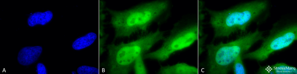
Immunocytochemistry/Immunofluorescence analysis using Rat Anti-HSF1 Monoclonal Antibody, Clone 10H8 (SMC-118). Tissue: Heat Shocked cervical cancer cells (HeLa). Species: Human. Fixation: 2% Formaldehyde for 20 min at RT. Primary Antibody: Rat Anti-HSF1 Monoclonal Antibody (SMC-118) at 1:100 for 12 hours at 4°C. Secondary Antibody: FITC Goat Anti-Rat (green) at 1:200 for 2 hours at RT. Counterstain: DAPI (blue) nuclear stain at 1:40000 for 2 hours at RT. Localization: Diffuse nuclear and cytoplasmic staining. Magnification: 100x. (A) DAPI (blue) nuclear stain. (B) Anti-HSF1 Antibody. (C) Composite. Heat Shocked at 42°C for 1h.
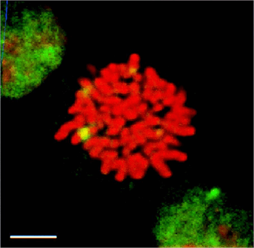
Immunocytochemistry/Immunofluorescence analysis using Rat Anti-HSF1 Monoclonal Antibody, Clone 10H8 (SMC-118). Tissue: Heat Shocked mitotic HeLa cells. Species: Human. Primary Antibody: Rat Anti-HSF1 Monoclonal Antibody (SMC-118) at 1:1000. HSF1 stained green. Courtesy of: Morimoto Lab, Northwestern University, USA.
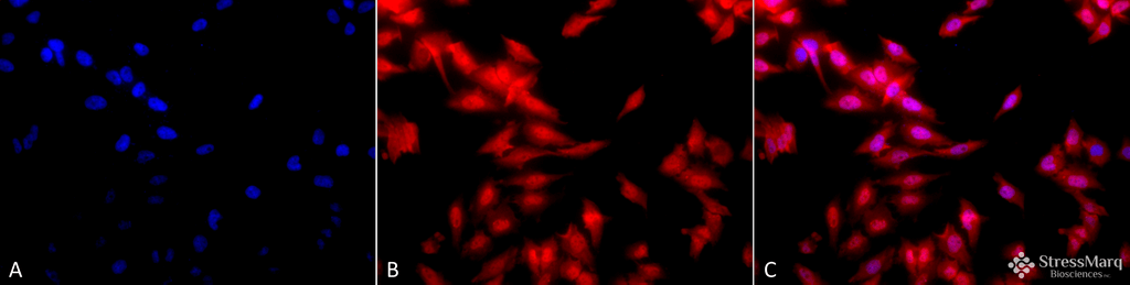
Immunocytochemistry/Immunofluorescence analysis using Rat Anti-HSF1 Monoclonal Antibody, Clone 10H8 (SMC-118). Tissue: Heat Shocked cervical cancer cells (HeLa). Species: Human. Fixation: 2% Formaldehyde for 20 min at RT. Primary Antibody: Rat Anti-HSF1 Monoclonal Antibody (SMC-118) at 1:100 for 12 hours at 4°C. Secondary Antibody: APC Goat Anti-Rat (red) at 1:200 for 2 hours at RT. Counterstain: DAPI (blue) nuclear stain at 1:40000 for 2 hours at RT. Localization: Diffuse nuclear and cytoplasmic staining. Magnification: 20x. (A) DAPI (blue) nuclear stain. (B) Anti-HSF1 Antibody. (C) Composite. Heat Shocked at 42°C for 1h.
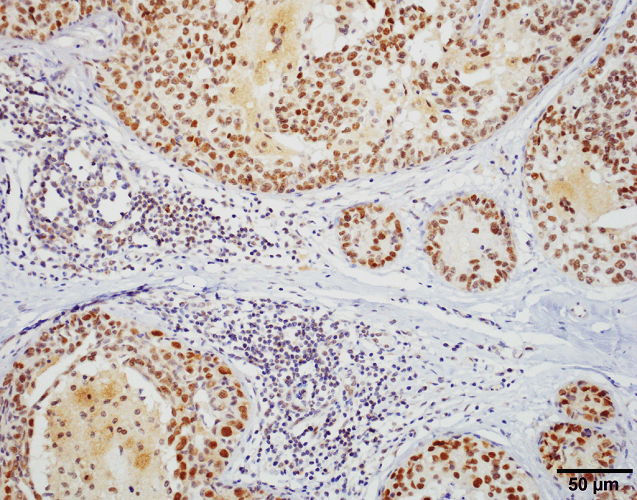
Immunohistochemistry analysis using Rat Anti-HSF1 Monoclonal Antibody, Clone 10H8 (SMC-118). Tissue: Breast carcinoma. Species: Human. Fixation: 10% Formalin Solution for 20 hours at RT. Primary Antibody: Rat Anti-HSF1 Monoclonal Antibody (SMC-118) at 1:1000 for 40 min. Secondary Antibody: Dako labeled Polymer HRP Anti-rat IgG, DAB Chromogen (brown) (Dako Envision+ System) for 30 min at RT. Counterstain: Mayer’s Hematoxylin (purple/blue) nuclear stain for 1 minute at RT. Localization: Nuclear. Magnification: 100X. Courtesy of: Dr. Sandro Santagata, Harvard Medical School.
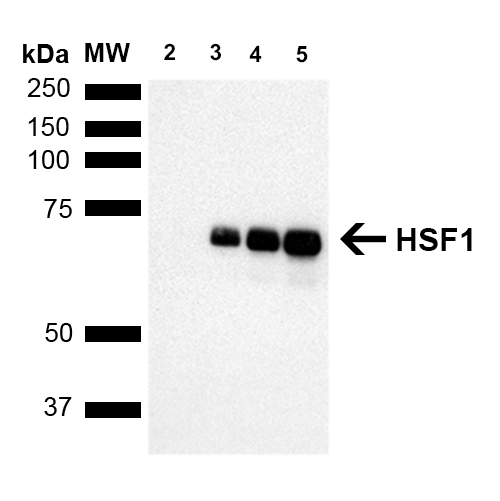
Western Blot analysis of Human Breast adenocarcinoma cell line (MCF7) showing detection of ~65 kDa HSF1 protein using Rat Anti-HSF1 Monoclonal Antibody, Clone 10H8 (SMC-118). Lane 1: MW ladder. Lane 2: HSF1 null lysate prepared from mouse embryonic fibroblasts. Lane 3: MCF7 lysate (5 µg). Lane 4: MCF7 lysate (10 µg). Lane 5: MCF7 lysate (20 µg). Block: 1.5% BSA for 30 minutes at RT. Primary Antibody: Rat Anti-HSF1 Monoclonal Antibody (SMC-118) at 1:1000 for 2 hours at RT. Secondary Antibody: Goat Anti-Rat IgG: HRP for 1 hour at RT. Predicted/Observed Size: ~65 kDa. Courtesy of: Dr. Sandro Santagata, Harvard Medical School.
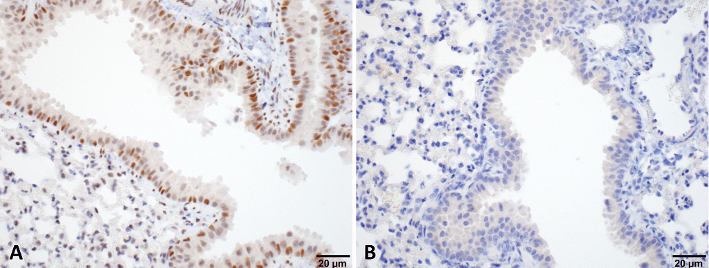
Immunohistochemistry analysis using Rat Anti-HSF1 Monoclonal Antibody, Clone 10H8 (SMC-118). Tissue: Lung. Species: Mouse. Fixation: 10% Formalin Solution for 20 hours at RT. Primary Antibody: Rat Anti-HSF1 Monoclonal Antibody (SMC-118) at 1:1000 for 40 min. Secondary Antibody: Dako labeled Polymer HRP Anti-rat IgG, DAB Chromogen (brown) (Dako Envision+ System) for 30 min at RT. Counterstain: Mayer’s Hematoxylin (purple/blue) nuclear stain for 1 minute at RT. Localization: Nuclear. Magnification: 100X. (A) HSF Wildtype. (B) HSF null. Courtesy of: Dr. Sandro Santagata, Harvard Medical School.
 Powered by Bioz
StressMarq Biosciences :
Based on validation through cited publications.