Advancing the Frontiers of Neurodegenerative Disease Research


Properties
| Storage Buffer | PBS pH7.4, 50% glycerol, 0.09% sodium azide *Storage buffer may change when conjugated |
| Storage Temperature | -20ºC, Conjugated antibodies should be stored according to the product label |
| Shipping Temperature | Blue Ice or 4ºC |
| Purification | Protein G Purified |
| Clonality | Monoclonal |
| Clone Number | 1F12-A6 |
| Isotype | IgG1 Kappa |
| Specificity | Detects 32kDa. Does not cross-react with HO-2. |
| Cite This Product | HO-1 Antibody (StressMarq Biosciences | Victoria, BC CANADA, Catalog# SMC-131, RRID: AB_2264116) |
| Certificate of Analysis | 1 µg/ml was sufficient for detection of HO-1 in 10 µg of mixed human cell line lysate by colorimetric immunoblot analysis using Goat Anti-Mouse IgG:HRP as the secondary. |
Biological Description
| Alternative Names | HSP32, HMOX1, Heme oxygenase 1, HO, HO1, 32 kD, bK286B10, D8Wsu38e, heat shock protein 32 kD, heat shock protein 32kD, Heat shock protein, Heme oxygenase (decycling) 1, Hemox, HMOX 1, Hmox, Hmox1, HMOX1_HUMAN, HO 1, HO-1, Heme Oxygenase-1, Haem Oxygenase-1, Heat Shock Protein 32, Heme Oxygenase (Decyclizing), EC 1.14.14.18 |
| Research Areas | Alzheimer's Disease, Atherosclerosis, Blood, Cancer, Cancer Metabolism, Cardiovascular System, Cell Signaling, Endothelium, Epigenetics and Nuclear Signaling, Hypoxia, Inflammatory Mediators, Metabolism, Metabolism processes, Neurodegeneration, Neuroscience, NFkB Pathway, Nuclear Signaling Pathways, Oxidative Stress, Platelets, Response to Hypoxia, Vascular Inflammation, Vasculature |
| Cellular Localization | Endoplasmic Reticulum, Microsome |
| Accession Number | NP_002124.1 |
| Gene ID | 3162 |
| Swiss Prot | P09601 |
| Scientific Background |
Heme oxygenase-1 (HO-1), also known as heat shock protein 32, is an inducible enzyme that catalyzes the degradation of heme into biliverdin, free iron, and carbon monoxide—each with distinct biological functions. Biliverdin is rapidly converted to bilirubin, a potent antioxidant, while carbon monoxide acts as a signaling molecule with anti-inflammatory and vasodilatory properties. Free iron, although potentially toxic, is sequestered by ferritin to limit oxidative damage. HO-1 is strongly upregulated in response to oxidative stress, inflammation, and neurotoxic insults, positioning it as a key cytoprotective factor in the central nervous system. Its expression is elevated in various neurodegenerative conditions, including Alzheimer’s disease, Parkinson’s disease, and multiple sclerosis, where it helps mitigate neuronal injury by reducing reactive oxygen species and modulating immune responses. Unlike its constitutive isoform HO-2, HO-1 is dynamically regulated and serves as a frontline defense against cellular stress. Deficiency in HO-1 has been linked to increased neuroinflammation, impaired stress tolerance, and heightened vulnerability to neurodegenerative pathology. Given its dual role in detoxifying free heme and generating neuroprotective metabolites, HO-1 is a promising therapeutic target for modulating oxidative stress and inflammation in neurodegenerative disease research. |
| References |
1. Froh M. et al. (2007) World J. Gastroentereol 13(25): 3478-86. 2. Elbirt K.K. and Bonkovsky H.L. (1999) Proc Assoc Am Physicians 111(5): 348-47. 3. Maines M.D., Trakshel G.M., and Kutty R.K. (1986) J Biol Chem 261: 411–419. 4. Brydun A., et al. (2007) Hypertens Res 30(4): 341-8. 5. Poss K.D. and Tonegawa S. (1997). Proc Natl Acad Sci U S A. 94: 10925–10930. 6. Yet S.F., et al. (2003) FASEB J. 17: 1759–1761. 7. Yachie A., et al. (1999) J Clin Invest. 103: 129–135. |
Product Images
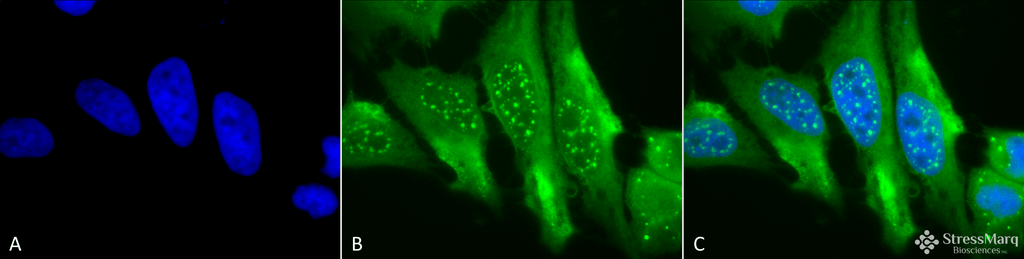
Immunocytochemistry/Immunofluorescence analysis using Mouse Anti-HO-1 Monoclonal Antibody, Clone 1F12-A6 (SMC-131). Tissue: Cervical cancer cell line (HeLa). Species: Human. Fixation: 2% Formaldehyde for 20 min at RT. Primary Antibody: Mouse Anti-HO-1 Monoclonal Antibody (SMC-131) at 1:100 for 12 hours at 4°C. Secondary Antibody: FITC Goat Anti-Mouse (green) at 1:200 for 2 hours at RT. Counterstain: DAPI (blue) nuclear stain at 1:40000 for 2 hours at RT. Localization: Microsome. Endoplasmic reticulum. Localizes to the nucleus upon hypoxia. Magnification: 100x. (A) DAPI (blue) nuclear stain. (B) Anti-HO-1 Antibody. (C) Composite.
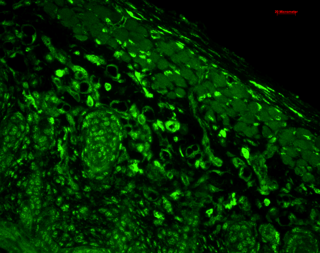
Immunohistochemistry analysis using Mouse Anti-HO-1 Monoclonal Antibody, Clone 1F12-A6 (SMC-131). Tissue: backskin. Species: Mouse. Fixation: Bouin’s Fixative and paraffin-embedded. Primary Antibody: Mouse Anti-HO-1 Monoclonal Antibody (SMC-131) at 1:100 for 1 hour at RT. Secondary Antibody: FITC Goat Anti-Mouse (green) at 1:50 for 1 hour at RT. Localization: muscle, dermis, hair follicles, epidermis: nuclear everywhere and some cytoplasmic staining.
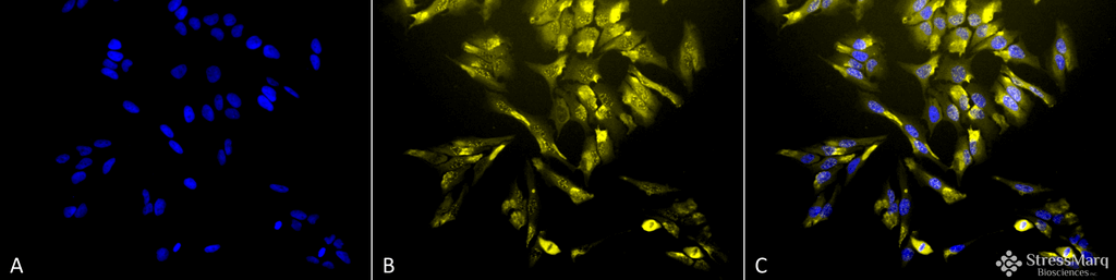
Immunocytochemistry/Immunofluorescence analysis using Mouse Anti-HO-1 Monoclonal Antibody, Clone 1F12-A6 (SMC-131). Tissue: Cervical cancer cell line (HeLa). Species: Human. Fixation: 2% Formaldehyde for 20 min at RT. Primary Antibody: Mouse Anti-HO-1 Monoclonal Antibody (SMC-131) at 1:100 for 12 hours at 4°C. Secondary Antibody: R-PE Goat Anti-Mouse (yellow) at 1:200 for 2 hours at RT. Counterstain: DAPI (blue) nuclear stain at 1:40000 for 2 hours at RT. Localization: Microsome. Endoplasmic reticulum. Localizes to the nucleus upon hypoxia. Magnification: 20x. (A) DAPI (blue) nuclear stain. (B) Anti-HO-1 Antibody. (C) Composite.

Western Blot analysis of Human Cervical cancer cell line (HeLa) lysate showing detection of HO-1 protein using Mouse Anti-HO-1 Monoclonal Antibody, Clone 1F12-A6 (SMC-131). Load: 15 µg. Block: 1.5% BSA for 30 minutes at RT. Primary Antibody: Mouse Anti-HO-1 Monoclonal Antibody (SMC-131) at 1:1000 for 2 hours at RT. Secondary Antibody: Sheep Anti-Mouse IgG: HRP for 1 hour at RT.
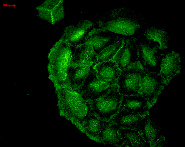
Immunocytochemistry/Immunofluorescence analysis using Mouse Anti-HO-1 Monoclonal Antibody, Clone 1F12-A6 (SMC-131). Tissue: HaCaT cells. Species: Human. Fixation: Cold 100% methanol for 10 minutes at -20°C. Primary Antibody: Mouse Anti-HO-1 Monoclonal Antibody (SMC-131) at 1:100 for 1 hour at RT. Secondary Antibody: FITC Goat Anti-Mouse (green) at 1:50 for 1 hour at RT. Localization: Cell-cell border staining in epidermis, punctuate nuclear staining. .
 Powered by Bioz
StressMarq Biosciences :
Based on validation through cited publications.