Discovery through Partnership | Excellence through Quality


Properties
| Storage Buffer | PBS pH7.4, 50% glycerol, 0.09% sodium azide *Storage buffer may change when conjugated |
| Storage Temperature | -20ºC, Conjugated antibodies should be stored according to the product label |
| Shipping Temperature | Blue Ice or 4ºC |
| Purification | Protein G Purified |
| Clonality | Recombinant Monoclonal |
| Clone Number | S70 |
| Isotype | IgG1 |
| Specificity | Detects ~100kDa. No cross-reactivity against HCN2. |
| Cite This Product | HCN1 Antibody (StressMarq Biosciences | Victoria, BC CANADA, Catalog# SMC-304, RRID: AB_2279461) |
| Certificate of Analysis | 1 µg/ml of SMC-304 was sufficient for detection of HCN1 in 10 µg of rat brain lysate by colorimetric immunoblot analysis using Goat anti-mouse IgG:HRP as the secondary antibody. |
Biological Description
| Alternative Names | BCNG-1, BCNG1, Brain cyclic nucleotide gated channel 1, Brain cyclic nucleotide-gated channel 1, HAC2, HCN1, HCN1_HUMAN, Hyperpolarization activated cyclic nucleotide gated potassium channel 1, Potassium/sodium hyperpolarization activated cyclic nucleotide gated channel 1, Potassium/sodium hyperpolarization-activated cyclic nucleotide-gated channel 1 |
| Research Areas | Cardiovascular System, Cyclic Nucleotide-Gated Ion Channels, Heart, Ion Channels, Neuroscience |
| Cellular Localization | Membrane |
| Accession Number | NP_445827.1 |
| Gene ID | 84390 |
| Swiss Prot | Q9JKB0 |
| Scientific Background |
Hyperpolarization-activated cyclic nucleotide-gated channel 1 (HCN1) is a voltage-gated ion channel that contributes to the generation and regulation of rhythmic electrical activity in neurons. It is predominantly expressed in the cortex, hippocampus, and cerebellum—regions critically involved in cognition, memory, and motor control. HCN1 channels play a key role in setting the resting membrane potential and modulating synaptic integration and neuronal excitability. In neurodegenerative diseases, such as Alzheimer’s and epilepsy-related cognitive decline, dysregulation of HCN1 expression or function has been linked to impaired synaptic plasticity and network dysfunction. Altered HCN1 activity can disrupt the balance of excitatory and inhibitory signaling, contributing to neuronal hyperexcitability and excitotoxicity—both of which are implicated in neurodegeneration. Moreover, HCN1 has been shown to influence dendritic integration and long-term potentiation (LTP), processes essential for learning and memory. Given its role in maintaining neuronal homeostasis and circuit stability, HCN1 is emerging as a potential therapeutic target for modulating brain excitability and protecting against neurodegenerative progression. |
| References | 1. Zong X., et al. (2005) J Biol Chem. 280(40): 34224-34232. |
Product Images
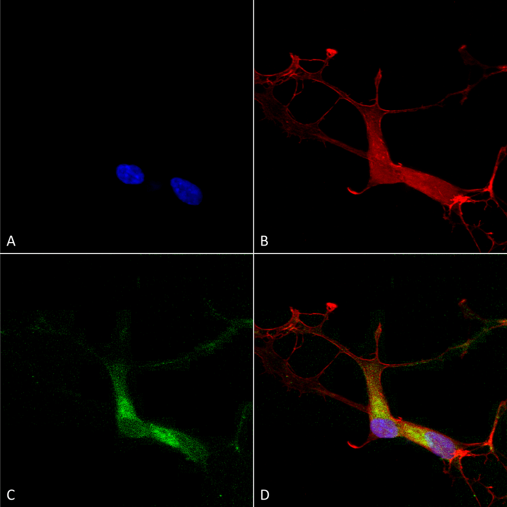
Immunocytochemistry/Immunofluorescence analysis using Mouse Anti-HCN1 Monoclonal Antibody, Clone S70 (SMC-304). Tissue: Neuroblastoma cells (SH-SY5Y). Species: Human. Fixation: 4% PFA for 15 min. Primary Antibody: Mouse Anti-HCN1 Monoclonal Antibody (SMC-304) at 1:100 for overnight at 4°C with slow rocking. Secondary Antibody: AlexaFluor 488 at 1:1000 for 1 hour at RT. Counterstain: Phalloidin-iFluor 647 (red) F-Actin stain; Hoechst (blue) nuclear stain at 1:800, 1.6mM for 20 min at RT. (A) Hoechst (blue) nuclear stain. (B) Phalloidin-iFluor 647 (red) F-Actin stain. (C) HCN1 Antibody (D) Composite.
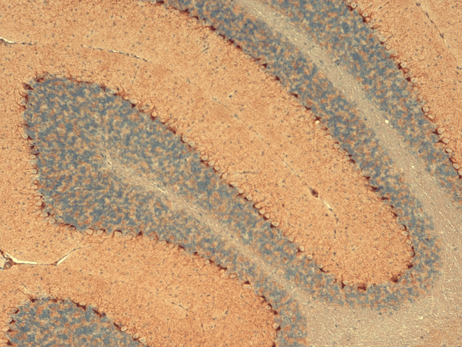
Immunohistochemistry analysis using Mouse Anti-HCN1 Monoclonal Antibody, Clone S70 (SMC-304). Tissue: Cerebellum. Species: Mouse. Fixation: 10% Formalin Solution for 12-24 hours at RT. Primary Antibody: Mouse Anti-HCN1 Monoclonal Antibody (SMC-304) at 1:1000 for 1 hour at RT. Secondary Antibody: HRP/DAB Detection System: Biotinylated Goat Anti-Mouse, Streptavidin Peroxidase, DAB Chromogen (brown) for 30 minutes at RT. Counterstain: Mayer Hematoxylin (purple/blue) nuclear stain at 250-500 µl for 5 minutes at RT. Localization: Cytoplasmic staining of Purkinje cells.
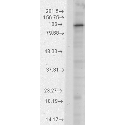
Western Blot analysis of Rat brain membrane lysate showing detection of HCN1 protein using Mouse Anti-HCN1 Monoclonal Antibody, Clone S70 (SMC-304). Load: 15 µg. Block: 1.5% BSA for 30 minutes at RT. Primary Antibody: Mouse Anti-HCN1 Monoclonal Antibody (SMC-304) at 1:1000 for 2 hours at RT. Secondary Antibody: Sheep Anti-Mouse IgG: HRP for 1 hour at RT.
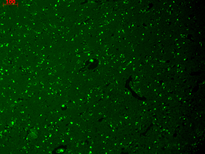
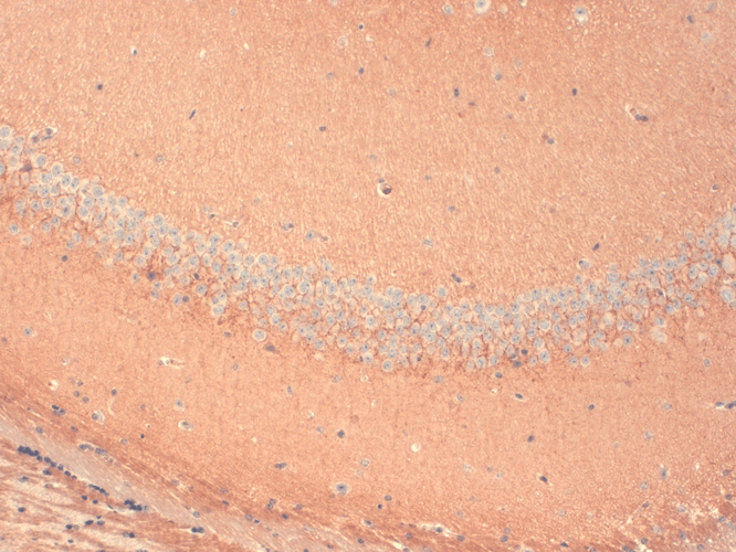
Immunohistochemistry analysis using Mouse Anti-HCN1 Monoclonal Antibody, Clone S70 (SMC-304). Tissue: hippocampus. Species: Human. Fixation: Bouin’s Fixative and paraffin-embedded. Primary Antibody: Mouse Anti-HCN1 Monoclonal Antibody (SMC-304) at 1:1000 for 1 hour at RT. Secondary Antibody: FITC Goat Anti-Mouse (green) at 1:50 for 1 hour at RT.
Immunohistochemistry analysis using Mouse Anti-HCN1 Monoclonal Antibody, Clone S70 (SMC-304). Tissue: Frozen brain section. Species: Mouse. Fixation: 10% Formalin Solution for 12-24 hours at RT. Primary Antibody: Mouse Anti-HCN1 Monoclonal Antibody (SMC-304) at 1:1000 for 1 hour at RT. Secondary Antibody: HRP/DAB Detection System: Biotinylated Goat Anti-Mouse, Streptavidin Peroxidase, DAB Chromogen (brown) for 30 minutes at RT. Counterstain: Mayer Hematoxylin (purple/blue) nuclear stain at 250-500 µl for 5 minutes at RT. Localization: Neurons.
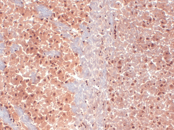
Immunohistochemistry analysis using Mouse Anti-HCN1 Monoclonal Antibody, Clone S70 (SMC-304). Tissue: Frozen brain section. Species: Mouse. Fixation: 10% Formalin Solution for 12-24 hours at RT. Primary Antibody: Mouse Anti-HCN1 Monoclonal Antibody (SMC-304) at 1:1000 for 1 hour at RT. Secondary Antibody: HRP/DAB Detection System: Biotinylated Goat Anti-Mouse, Streptavidin Peroxidase, DAB Chromogen (brown) for 30 minutes at RT. Counterstain: Mayer Hematoxylin (purple/blue) nuclear stain at 250-500 µl for 5 minutes at RT.
 Powered by Bioz
StressMarq Biosciences :
Based on validation through cited publications.