Properties
| Storage Buffer | PBS pH7.4, 50% glycerol, 0.09% sodium azide *Storage buffer may change when conjugated |
| Storage Temperature | -20ºC, Conjugated antibodies should be stored according to the product label |
| Shipping Temperature | Blue Ice or 4ºC |
| Purification | Protein G Purified |
| Clonality | Monoclonal |
| Clone Number | N330A/80 (Formerly sold as S330A-80) |
| Isotype | IgG1 |
| Specificity | Detects 50kDa or larger (possibly due to dimerization). Does not cross-react with other adrenergic receptors. |
| Cite This Product | Alpha-2C Adrenergic Receptor Antibody (StressMarq Biosciences | Victoria, BC CANADA, Catalog# SMC-435, RRID: AB_2701577) |
| Certificate of Analysis | 1 µg/ml of SMC-435 was sufficient for detection of Alpha2C Adrenergic Receptor in 20 µg of COS cells transiently transfected with HA-tagged Alpha- lysate and assayed by colorimetric immunoblot analysis using goat anti-mouse IgG:HRP as the secondary antibody. |
Biological Description
| Alternative Names | Alpha-2C Adrenergic Receptor, Alpha-2C Adrenoceptor, Alpha-2C Adrenoreceptor, ADRA2C, ADRA2L2, ADRA2RL2, ADRARL2, ALPHA2CAR, Alpha2-AR-C4, Alpha2-C4, Adrenoceptor Alpha 2C, Adrenergic receptor alpha 2C |
| Research Areas | Alpha-2 Adrenergic Receptors, Cell Signaling, Neuroscience, Neurotransmitter Receptors |
| Cellular Localization | Cell membrane |
| Accession Number | NP_000674.2 |
| Gene ID | 152 |
| Swiss Prot | P18825 |
| Scientific Background |
The Alpha-2C Adrenergic Receptor (ADRA2C) is a G protein-coupled receptor that plays a critical role in modulating neurotransmitter release from central adrenergic neurons and sympathetic nerves, particularly in the brain and heart. Widely expressed in the central nervous system, ADRA2C is involved in regulating synaptic transmission, stress responses, and higher-order cognitive and behavioral functions. Two isoforms of ADRA2C are produced via alternative splicing, with predominant expression in brain regions implicated in memory, emotion, and executive function. Beyond the CNS, ADRA2C is also found in peripheral tissues including the heart, kidney, blood, and corpus cavernosum, suggesting a broader physiological role. In the context of neurodegenerative disease, dysregulation of ADRA2C signaling has been linked to impaired noradrenergic tone, altered synaptic plasticity, and neuroinflammation—key features observed in disorders such as Alzheimer’s disease, Parkinson’s disease, and schizophrenia. Its involvement in modulating cognitive resilience and behavioral regulation positions ADRA2C as a promising biomarker and therapeutic target in neuropsychiatric and neurodegenerative research. Ongoing studies are exploring ADRA2C agonists and antagonists for their potential to restore neurotransmitter balance, enhance cognitive function, and mitigate neurodegenerative progression |
| References |
1. Filipeanu C.M., et al. (2011) Biochim Biophys Acta. 1813: 346-357. 2. Powe D.G., et al. (2011) Breast Cancer Res Treat. 130: 457-463. |
Product Images

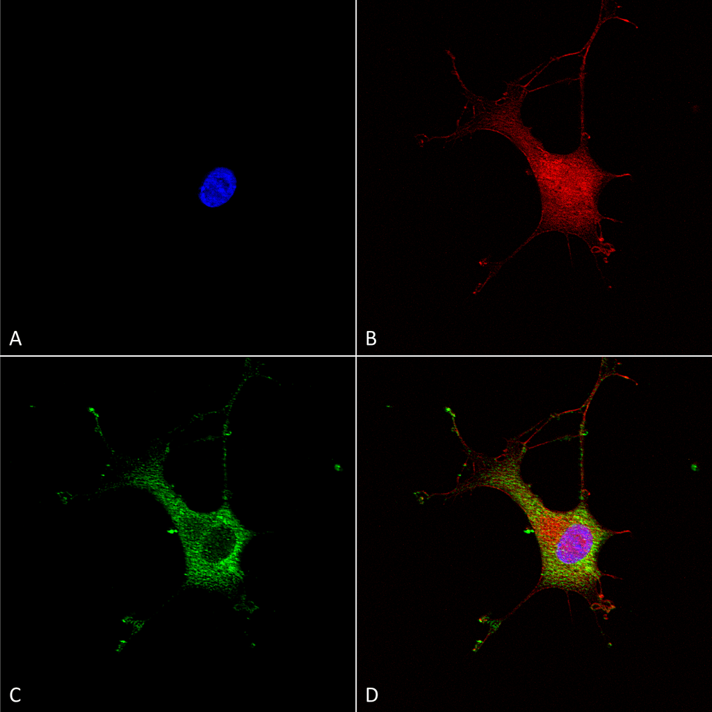
Immunocytochemistry/Immunofluorescence analysis using Mouse Anti-Alpha-2C Adrenergic Receptor Monoclonal Antibody, Clone N330A/80 (SMC-435). Tissue: Neuroblastoma cells (SH-SY5Y). Species: Human. Fixation: 4% PFA for 15 min. Primary Antibody: Mouse Anti-Alpha-2C Adrenergic Receptor Monoclonal Antibody (SMC-435) at 1:100 for overnight at 4°C with slow rocking. Secondary Antibody: AlexaFluor 488 at 1:1000 for 1 hour at RT. Counterstain: Phalloidin-iFluor 647 (red) F-Actin stain; Hoechst (blue) nuclear stain at 1:800, 1.6mM for 20 min at RT. (A) Hoechst (blue) nuclear stain. (B) Phalloidin-iFluor 647 (red) F-Actin stain. (C) Alpha-2C Adrenergic Receptor Antibody (D) Composite.

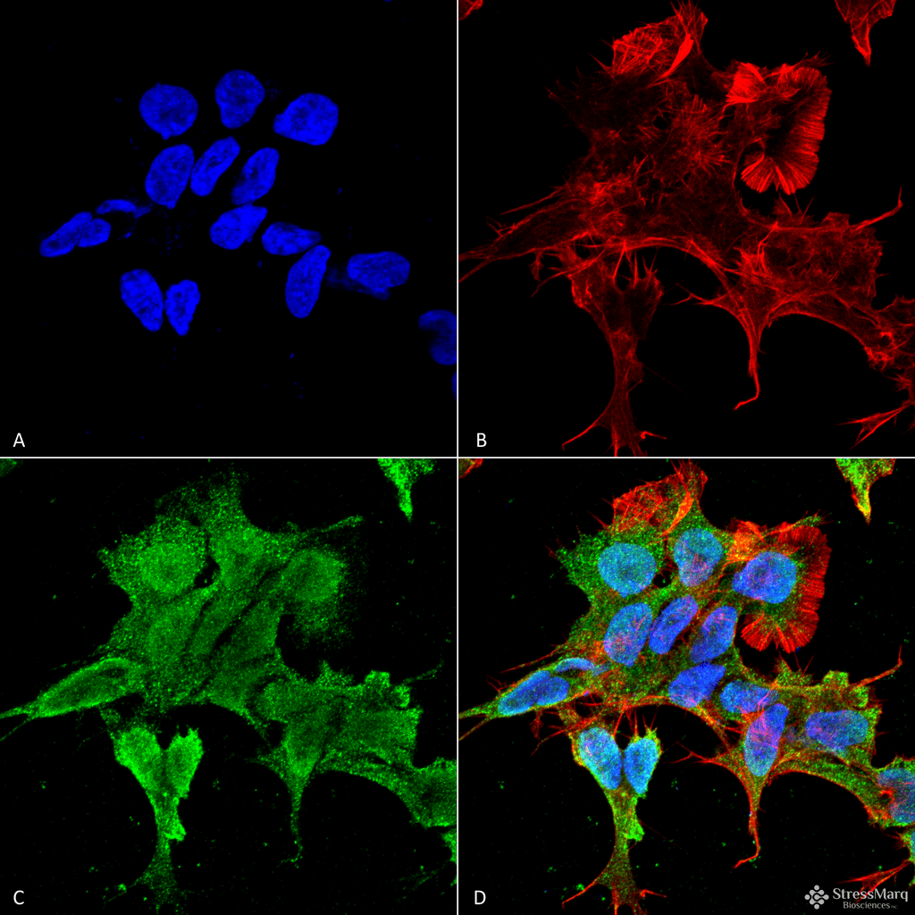
Immunocytochemistry/Immunofluorescence analysis using Mouse Anti-Alpha-2C Adrenergic Receptor Monoclonal Antibody, Clone N330A/80 (SMC-435). Tissue: Neuroblastoma cell line (SK-N-BE). Species: Human. Fixation: 4% Formaldehyde for 15 min at RT. Primary Antibody: Mouse Anti-Alpha-2C Adrenergic Receptor Monoclonal Antibody (SMC-435) at 1:100 for 60 min at RT. Secondary Antibody: Goat Anti-Mouse ATTO 488 at 1:100 for 60 min at RT. Counterstain: Phalloidin Texas Red F-Actin stain; DAPI (blue) nuclear stain at 1:1000, 1:5000 for 60min RT, 5min RT. Localization: Cell Membrane, Nucleus. Magnification: 60X. (A) DAPI (blue) nuclear stain. (B) Phalloidin Texas Red F-Actin stain. (C) Alpha-2C Adrenergic Receptor Antibody. (D) Composite.

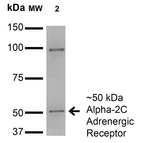
Western Blot analysis of Monkey COS cells transfected with HA-tagged Alpha-2C showing detection of ~50 kDa Alpha-2C Adrenergic Receptor protein using Mouse Anti-Alpha-2C Adrenergic Receptor Monoclonal Antibody, Clone N330A/80 (SMC-435). Lane 1: Molecular Weight Ladder. Lane 2: Monkey COS cells transfected with HA-tagged Alpha-2C. Load: 15 µg. Block: 2% BSA and 2% Skim Milk in 1X TBST. Primary Antibody: Mouse Anti-Alpha-2C Adrenergic Receptor Monoclonal Antibody (SMC-435) at 1:200 for 16 hours at 4°C. Secondary Antibody: Goat Anti-Mouse IgG: HRP at 1:1000 for 1 hour RT. Color Development: ECL solution for 6 min in RT. Predicted/Observed Size: ~50 kDa.


Reviews
There are no reviews yet.