Advancing the Frontiers of Neurodegenerative Disease Research


Properties
| Storage Buffer | PBS pH 7.4, 50% glycerol, 0.09% sodium azide *Storage buffer may change when conjugated |
| Storage Temperature | -20ºC, Conjugated antibodies should be stored according to the product label |
| Shipping Temperature | Blue Ice or 4ºC |
| Purification | Peptide Affinity Purified |
| Clonality | Polyclonal |
| Specificity | Binds to phosphorylated serine 129 on alpha synuclein. Does not detect unphosphorylated serine 129 alpha synuclein. Detects bands at 100, 75, 45, 15 kDa. Bands above 15 kDa are oligomers. |
| Cite This Product | Alpha Synuclein Antibody (pSer129) (StressMarq Biosciences | Victoria, BC CANADA, Catalog# SPC-742, RRID: AB_2706527) |
| Certificate of Analysis | A 1:1000 dilution of SPC-742 was sufficient for detection of Alpha Synuclein pSer129 in 15 µg of human brain cell lysates by ECL immunoblot analysis using goat anti-rabbit IgG:HRP as the secondary antibody. |
Biological Description
| Alternative Names | Alpha Synuclein, α-Synuclein, SNCA, alphaSYN, NACP, Non-A beta component of AD amyloid, Non-A4 component of amyloid precursor, Phosphorylated alpha synuclein, Phospho-alpha Synuclein (S129), Alpha-synuclein (phospho S129), Alpha Synuclein (phospho Ser129), Alpha-Synuclein Phospho Ser129, phospho-α-Synuclein (Ser129), Alpha Synuclein phospho Serine 129, Alpha Synuclein phospho Ser 129, Alpha Synuclein pSerine 129, Alpha Synuclein pSer 129, Alpha Synuclein phosphoSer 129, isoform NACP140, PARK1, PARK 1, PARK4, PARK 4, Parkinson disease (autosomal dominant, Lewy body) 4, Parkinson disease familial 1, SYN, Synuclein alpha, Synuclein alpha 140, Synuclein, alpha (non A4 component of amyloid precursor), SYUA_HUMAN |
| Research Areas | Alzheimer's Disease, Neurodegeneration, Neuroscience, Parkinson's Disease, Synuclein, Tangles & Tau, Multiple System Atrophy |
| Cellular Localization | Cell Junction, Cytoplasm, Membrane, Nucleus, Secreted, Synapse |
| Accession Number | NP_000336.1 |
| Gene ID | 6622 |
| Swiss Prot | P37840 |
| Scientific Background |
Alpha-synuclein (SNCA) is a neuronal protein predominantly expressed in the brain, where it is enriched at presynaptic terminals and plays a key role in synaptic function (1). It is also highly expressed in mitochondria-rich regions such as the olfactory bulb, hippocampus, striatum, and thalamus, suggesting a role in mitochondrial dynamics and neuronal energy metabolism (2). Functionally, alpha-synuclein interacts with tubulin (3), indicating a potential role as a microtubule-associated protein involved in cytoskeletal organization and intracellular transport. It is essential for cognitive development, with SNCA inactivation linked to impaired spatial learning and working memory (4). Pathologically, SNCA aggregates are a major non-Aβ component of amyloid plaques in Alzheimer’s disease and a defining feature of Lewy body inclusions in Parkinson’s disease (PD). PD is characterized by the progressive accumulation of alpha-synuclein and ubiquitin-positive inclusions in vulnerable neurons, contributing to neurodegeneration (5, 6). Critically, phosphorylation of alpha-synuclein at serine 129 (pSer129) is a key post-translational modification associated with disease pathology. Approximately 90% of alpha-synuclein found in Lewy bodies is phosphorylated at Ser129, compared to only a small fraction in the healthy brain (7, 8). This modification is widely used as a biomarker for pathological alpha-synuclein and is a focal point in therapeutic and diagnostic research targeting synucleinopathies. |
| References |
1. “Genetics Home Reference: SNCA”. US National Library of Medicine. (2013). 2. Zhang L., et al. (2008) Brain Res. 1244: 40-52. 3. Alim M.A., et al. (2002) J Biol Chem. 277(3): 2112-2117. 4. Kokhan V.S., Afanasyeva M.A., Van'kin G. (2012) Behav. Brain. Res. 231(1): 226-230. 5. Spillantini M.G., et al. (1997) Nature. 388(6645): 839-840. 6. Mezey E., et al. (1998) Nat Med. 4(7): 755-757. 7. Fujiwara H., et al. (2002) Nat Cell Biol. 4(2):160-4. 8. Anderson J.P., et al. (2006) J Biol Chem. 281(40):29739-52. |
Product Images
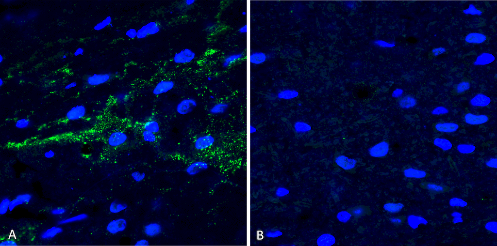
Immunohistochemistry analysis using Rabbit Anti-Alpha Synuclein (pSer129) Polyclonal Antibody (SPC-742). Tissue: Free floating brain sections. Species: Mouse. Fixation: PFA. Primary Antibody: Rabbit Anti-Alpha Synuclein (pSer129) Polyclonal Antibody (SPC-742) at 1:500 for overnight at 4C with gentle agitation. Counterstain: Hoechst. Magnification: 63X. A) Right hemisphere (striatum) injected with alpha synuclein AAV vector. B) Control. Alpha synuclein streaks are visible at injection site, but not control, 4 months after injection. Courtesy of: Trine Rasmussen, Simon Molgaard Jensen, Aarhus University.
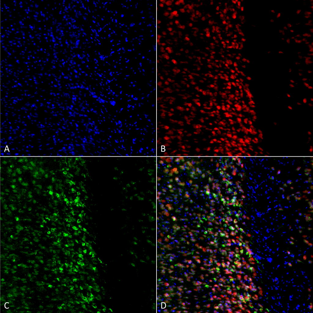
Immunocytochemistry/Immunofluorescence analysis using Rabbit Anti-Alpha Synuclein pSer129 Polyclonal Antibody (SPC-742). Tissue: Primary hippocampal neurons treated with active Alpha Synuclein Protein Aggregate (SPR-322) at 4 µg/ml to induce fibrils. Species: Rat. Fixation: 4% paraformaldehyde. Primary Antibody: Rabbit Anti-Alpha Synuclein pSer129 Polyclonal Antibody (SPC-742) at 1:500 for 24 hours at 4°C. Secondary Antibody: Goat Anti-Rabbit Alexa Fluor 488 at 1:700 for 1 hour at RT. Counterstain: Guinea Pig Anti-NeuN (red) neuronal marker (Donkey Anti-Guinea Pig Alexa Fluor 647 1:700); Hoechst (blue) nuclear stain at 1:6000, 1:3000 for 60 min at RT, 5 min at RT. Magnification: 20X.
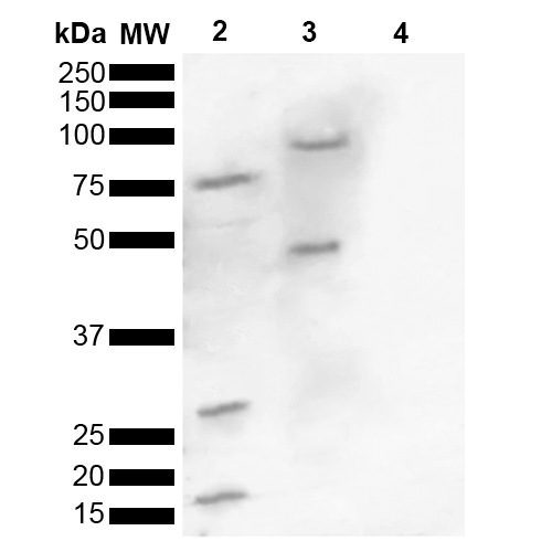
Western blot analysis of Human, Mouse brain lysate showing detection of ~16 kDa Alpha Synuclein pSer129 protein using Rabbit Anti-Alpha Synuclein pSer129 Polyclonal Antibody (SPC-742). Lane 1: Molecular Weight Ladder (MW). Lane 2: Human brain lysate. Lane 3: Mouse brain lysate. Lane 4: Human Alpha Synuclein Monomer (0.5 µg). Load: 15 µg. Block: 5% Skim Milk in 1X TBST. Primary Antibody: Rabbit Anti-Alpha Synuclein pSer129 Polyclonal Antibody (SPC-742) at 1:1000 for 2 hours at RT. Secondary Antibody: Goat Anti-Rabbit HRP:IgG at 1:3000 for 1 hour at RT. Color Development: ECL solution for 5 min at RT. Predicted/Observed Size: ~16 kDa. Other Band(s): 100, 75, 45, 30,16 kDa.
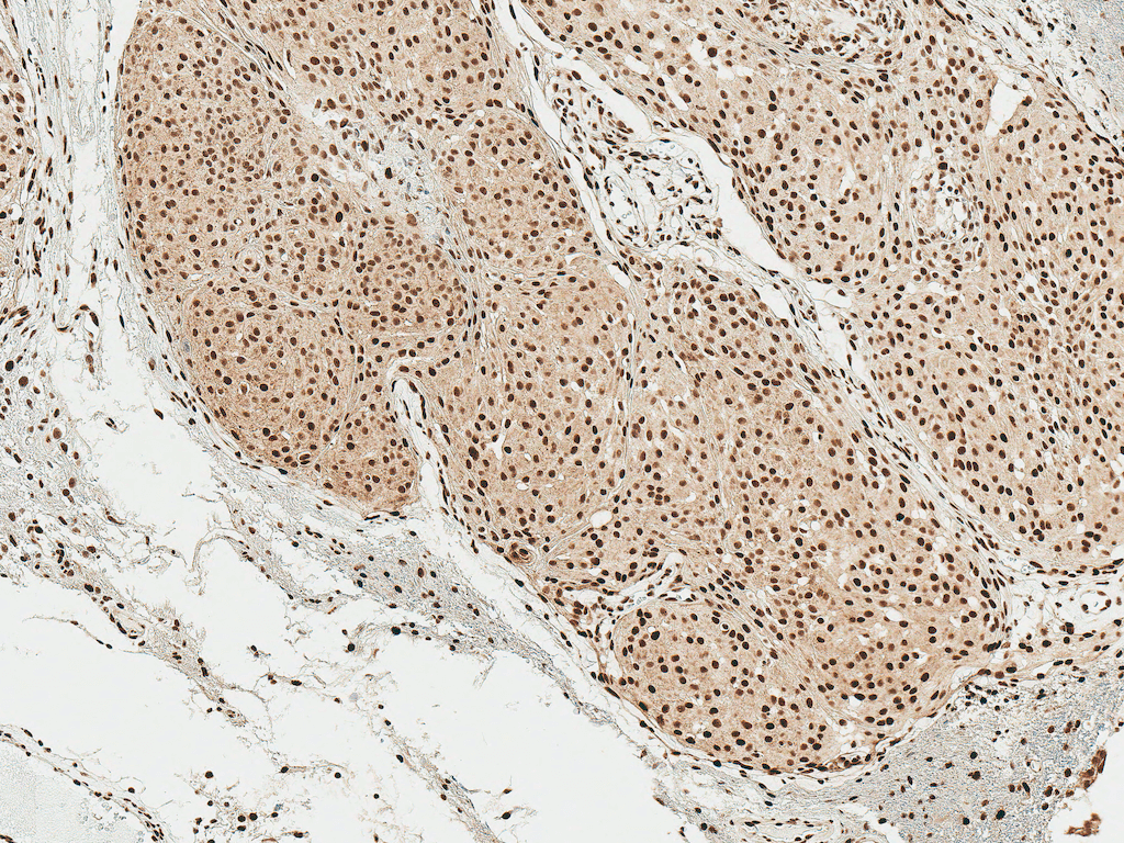
Immunohistochemistry analysis using Rabbit Anti-Alpha Synuclein pSer129 Polyclonal Antibody (SPC-742). Tissue: Brain. Species: Human. Fixation: Formalin Fixed Paraffin-Embedded. Primary Antibody: Rabbit Anti-Alpha Synuclein pSer129 Polyclonal Antibody (SPC-742) at 1:50 for 30 min at RT. Counterstain: Hematoxylin. Magnification: 10X. HRP-DAB Detection.
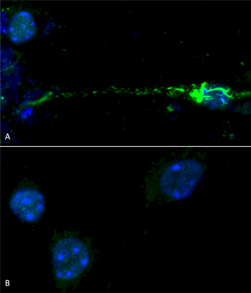
Immunocytochemistry/Immunofluorescence analysis using Rabbit Anti-Alpha Synuclein (pSer129) Polyclonal Antibody (SPC-742). Species: Mouse. Primary Antibody: Rabbit Anti-Alpha Synuclein (pSer129) Polyclonal Antibody (SPC-742). Phospho serine 129 antibody (SPC-742) was used to detect phosphorylated alpha synuclein in primary mouse hippocampal neurons treated with 100 nM sonicated mouse alpha synuclein PFFs (SPR-324) (A). Phosphorylated alpha synuclein was visible in perinucleus and neurites compared to untreated control (B). Read the protocol at pabmabs.com/?p=7901. Image courtesy of Trine Rasmussen, Simon Molgaard Jensen at Aarhus University..
 Powered by Bioz
Trine Rasmussen :
Read the full review on pAbmAbs.com“””