Discovery through Partnership | Excellence through Quality


Properties
| Storage Buffer | PBS, 50% glycerol, 0.09% sodium azide *Storage buffer may change when conjugated |
| Storage Temperature | -20ºC, Conjugated antibodies should be stored according to the product label |
| Shipping Temperature | Blue Ice or 4ºC |
| Purification | Peptide Affinity Purified |
| Clonality | Polyclonal |
| Specificity | Detects 77.9 kDa. |
| Cite This Product | ATG7 Antibody (StressMarq Biosciences | Victoria, BC CANADA, Catalog# SPC-610, RRID: AB_2704889) |
| Certificate of Analysis | A 1:1000 dilution of SPC-610 was sufficient for detection of ATG7 in 15 µg of human HeLa cell lysates by ECL immunoblot analysis using goat anti-rabbit IgG:HRP as the secondary antibody. |
Biological Description
| Alternative Names | ATG7, ATG7_HUMAN, APG7, APG7L, APG7-like, APG7 autophagy 7 like, APG7 autophagy 7-like (S. cerevisiae), ATG7 autophagy related 7 homolog, ATG7 autophagy related 7 homolog (S. cerevisiae), ATG12-activating enzyme E1 ATG7, Ubiquitin-like modifier-activating enzyme ATG7, Ubiquitin-activating enzyme E1-like protein, Ubiquitin activating enzyme E1 like protein, Autophagy-related protein 7, Autophagy related protein 7, Autophagy-related 7 (yeast), Autophagy 7 S. cerevisiae homolog of, GSA7, GSA 7, hAGP7, Atg7l, DKFZp434N0735, 1810013K23Rik, ATG 7, Apg 7 |
| Research Areas | Apoptosis, Autophagy, Cancer, Cardiovascular System, Heart |
| Cellular Localization | Cytoplasm, Preautophagosomal structure |
| Accession Number | NP_001129503.2 |
| Gene ID | 10533 |
| Swiss Prot | O95352 |
| Scientific Background |
ATG7 is a pivotal E1-like enzyme essential for autophagosome biogenesis, a core process in cellular homeostasis and neuroprotection. Functioning in concert with ATG10, ATG7 catalyzes the conjugation of ATG12 to ATG5, a critical step in the formation of the autophagosome membrane. Additionally, ATG7 activates ATG8 (LC3/GABARAP family proteins), facilitating their lipidation and incorporation into autophagosomal membranes. This dual role positions ATG7 as a master regulator of autophagic flux. In the context of neuroscience, ATG7 has emerged as a key player in the maintenance of neuronal integrity. Its activity is vital for the clearance of misfolded proteins and damaged organelles—hallmarks of neurodegenerative diseases such as Alzheimer's, Parkinson's, and Huntington's disease. Conditional knockout models have demonstrated that loss of ATG7 in neurons leads to progressive neurodegeneration, underscoring its neuroprotective function. Moreover, ATG7 is critical for metabolic adaptation, particularly in neonates, where it ensures amino acid supply through autophagy during nutrient scarcity. This metabolic role further highlights its importance in brain development and function. Given its central role in autophagy and neuronal survival, ATG7 is a promising therapeutic target in neurodegenerative disease research. Ongoing studies aim to modulate ATG7 activity to restore autophagic balance and mitigate neurodegeneration. |
| References |
1. Mizushima N., et al. (1998) J Biol Chem. 273: 33889-92. 2. Mizushima N., et al. (1998) Nature. 395: 395-8. 3. Suzuki K., et al. (2001) EMBO J. 20: 5971-81. 4. Tanida I., et al. (1999) Mol Biol Cell. 10: 1367-79. 5. Shintani T., et al. (1999) EMBO J. 18: 5234-41. |
Product Images
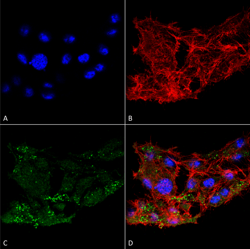
Immunocytochemistry/Immunofluorescence analysis using Rabbit Anti-ATG7 Polyclonal Antibody (SPC-610). Tissue: Colon carcinoma cell line (RKO). Species: Human. Fixation: 4% Formaldehyde for 15 min at RT. Primary Antibody: Rabbit Anti-ATG7 Polyclonal Antibody (SPC-610) at 1:100 for 60 min at RT. Secondary Antibody: Goat Anti-Rabbit ATTO 488 at 1:100 for 60 min at RT. Counterstain: Phalloidin Texas Red F-Actin stain; DAPI (blue) nuclear stain at 1:1000, 1:5000 for 60 min at RT, 5 min at RT. Localization: Cytoplasm, Preautophagosomal Structure, Organelle membrane. Magnification: 60X. (A) DAPI nuclear stain. (B) Phalloidin Texas Red F-Actin stain. (C) ATG7 Antibody. (D) Composite.
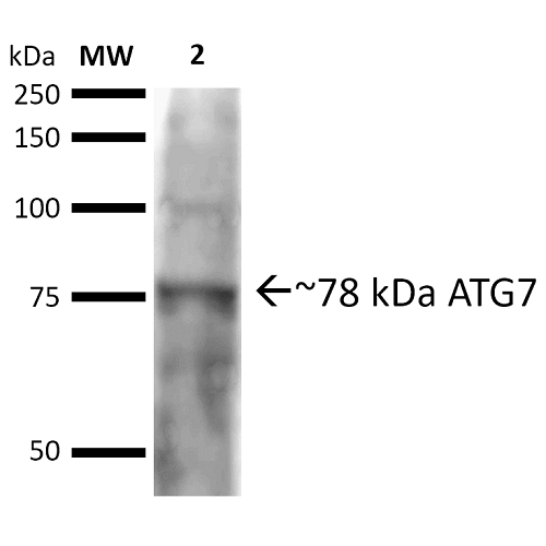
Western blot analysis of Rat brain cell lysates showing detection of ~77.9 kDa ATG7 protein using Rabbit Anti-ATG7 Polyclonal Antibody (SPC-610). Lane 1: Molecular Weight Ladder (MW). Lane 2: Rat brain cell lysates. Load: 20 µg. Block: 2% BSA and 2% Skim Milk in 1X TBST. Primary Antibody: Rabbit Anti-ATG7 Polyclonal Antibody (SPC-610) at 1:1000 for 16 hours at 4°C. Secondary Antibody: Goat Anti-Rabbit IgG: HRP at 1:2000 for 60 min at RT. Color Development: ECL solution for 6 min at RT. Predicted/Observed Size: ~77.9 kDa.
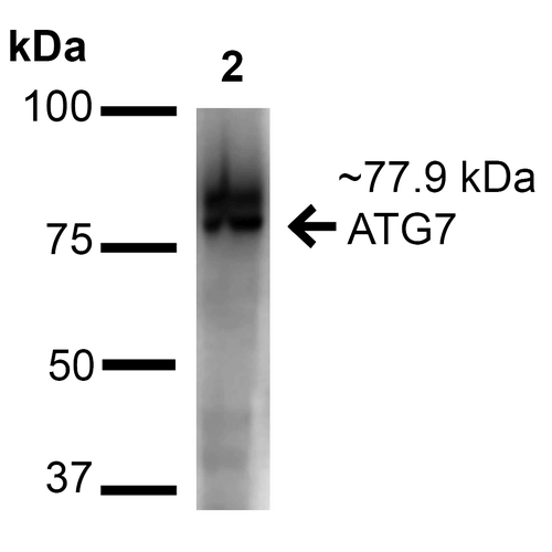
Western blot analysis of Human Cervical cancer cell line (HeLa) lysate showing detection of ~77.9 kDa ATG7 protein using Rabbit Anti-ATG7 Polyclonal Antibody (SPC-610). Lane 1: Molecular Weight Ladder (MW). Lane 2: Human Cervical cancer cell line (HeLa) lysate. Load: 15 µg. Block: 5% Skim Milk in 1X TBST. Primary Antibody: Rabbit Anti-ATG7 Polyclonal Antibody (SPC-610) at 1:1000 for 60 min at RT. Secondary Antibody: Goat Anti-Rabbit IgG: HRP at 1:2000 for 60 min at RT. Color Development: ECL solution for 6 min at RT. Predicted/Observed Size: ~77.9 kDa.
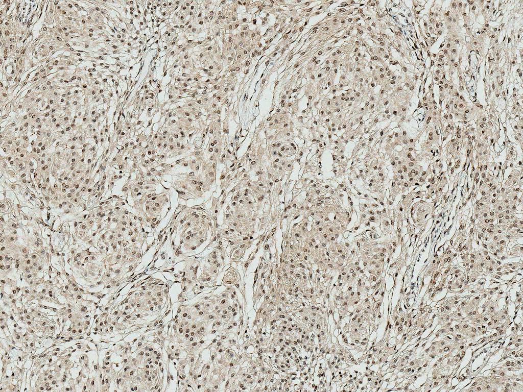
Immunohistochemistry analysis using Rabbit Anti-ATG10 Polyclonal Antibody (SPC-610). Tissue: Brain. Species: Human. Fixation: Formalin Fixed Paraffin-Embedded. Primary Antibody: Rabbit Anti-ATG10 Polyclonal Antibody (SPC-610) at 1:50 for 31 min at RT. Counterstain: Hematoxylin. Magnification: 10X.
 Powered by Bioz
Reviews
There are no reviews yet.