Advancing the Frontiers of Neurodegenerative Disease Research


Properties
| Storage Buffer | PBS pH7.4, 50% glycerol, 0.09% sodium azide *Storage buffer may change when conjugated |
| Storage Temperature | -20ºC, Conjugated antibodies should be stored according to the product label |
| Shipping Temperature | Blue Ice or 4ºC |
| Purification | Peptide Affinity Purified |
| Clonality | Polyclonal |
| Specificity | Detects ~63kDa. |
| Cite This Product | Calreticulin Antibody (StressMarq Biosciences | Victoria, BC CANADA, Catalog# SPC-122, RRID: AB_2069601) |
| Certificate of Analysis | A 1:10,000 dilution of SPC-122 was sufficient for detection of Calreticulin in 20 µg of HeLa cell lysate by ECL immunoblot analysis. |
Biological Description
| Alternative Names | Calreticulin, CALR, CALR_HUMAN, Calregulin, cC1qR, CRP55, ERp60, HSCBP, RO, SSA, grp60 |
| Research Areas | Cell Signaling, Organelle Markers, Organelle Proteins, Protein Trafficking, Tags and Cell Markers |
| Cellular Localization | Cell Surface, Cytoplasm, Endoplasmic Reticulum, Endoplasmic reticulum lumen, Extracellular Matrix, Sarcoplasmic Reticulum, Sarcoplasmic Reticulum Lumen |
| Accession Number | NP_004334.1 |
| Gene ID | 811 |
| Swiss Prot | P27797 |
| Scientific Background |
Calreticulin (CALR) is a highly conserved, multifunctional calcium-binding protein predominantly localized to the endoplasmic reticulum (ER), but also detected in the nucleus and nuclear envelope. It contains a C-terminal KDEL sequence that ensures ER retention, a hallmark of many ER-resident chaperones. Structurally, Calreticulin comprises three domains: a 180-residue N-terminal domain, a proline-rich P-domain (residues 189–288) with high-affinity calcium-binding capacity and homology to calnexin and calmegin, and a C-terminal domain that binds calcium with low affinity but high capacity. This architecture enables Calreticulin to function as both a calcium buffer and a molecular chaperone. In the nervous system, Calreticulin plays a critical role in protein folding, calcium signaling, and ER stress responses—processes intimately linked to neurodegenerative disease. It assists in the maturation of glycoproteins and regulates intracellular calcium dynamics, which are essential for synaptic function and neuronal survival. Dysregulation of Calreticulin has been implicated in neurodegenerative conditions such as Alzheimer’s disease, Parkinson’s disease, and amyotrophic lateral sclerosis (ALS), where ER stress and disrupted proteostasis are central pathological features. Beyond its chaperone role, Calreticulin influences gene expression, cytoskeletal organization, and cellular proliferation. It is also upregulated in response to various stressors, including amino acid deprivation, highlighting its role in adaptive cellular responses. Given its central position at the intersection of calcium homeostasis, protein quality control, and stress signaling, Calreticulin is a promising target in neurodegenerative disease research. |
| References |
1. Johnson S., et al. (2001) Trends Cell Biol 11: 122-129. 2. Smith M.J., et al. (1989) EMBO J. 8: 3581-3586. 3. Ellgaard L., et al. (2001) Curr Opin Cell Biol. 13: 431-437. 4. Krause K.H., and Michalak M. (1997) Cell. 88: 439-443. 5. Nash P.D., et al. (1994) Mol Cell Biochem. 135: 71-78. 6. Heal R., and McGivan J. (1998) Biochem J. 329: 389-394. 7. Lucero H.A., et al. (1998) J Biol Chem. 273: 9857-9863. 8. Tanaka S., et al. (2000) J Biol Chem. 275: 10388-10393. 9. Yoon G.S., et al. (2000) Cancer Res. 60: 1117-1120. 10. Antoniou A.N., et al. (2002) Immunology 106: 182-189. 11. Wada I., et al. (1997) EMBO J. 16: 5420-5432. 12. Laguerre D.B., et al. (1998) J Vir. 72: 4940-4949. |
Product Images
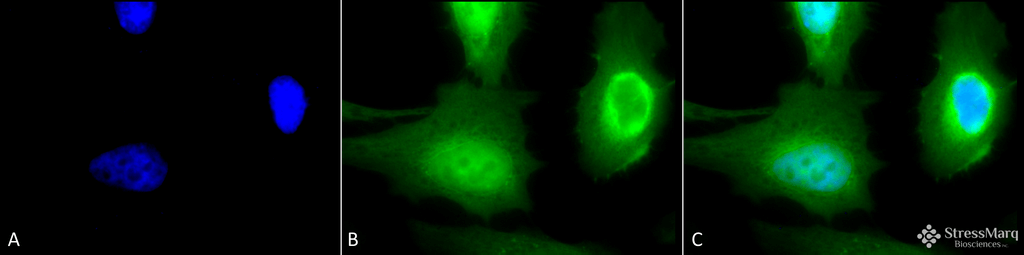
Immunocytochemistry/Immunofluorescence analysis using Rabbit Anti-Calreticulin Polyclonal Antibody (SPC-122). Tissue: Heat Shocked Cervical cancer cell line (HeLa). Species: Human. Fixation: 2% Formaldehyde for 20 min at RT. Primary Antibody: Rabbit Anti-Calreticulin Polyclonal Antibody (SPC-122) at 1:100 for 12 hours at 4°C. Secondary Antibody: FITC Goat Anti-Rabbit (green) at 1:200 for 2 hours at RT. Counterstain: DAPI (blue) nuclear stain at 1:40000 for 2 hours at RT. Localization: Endoplasmic reticulum lumen. Cytoplasm. Magnification: 100x. (A) DAPI (blue) nuclear stain. (B) Anti-Calreticulin Antibody. (C) Composite. Heat Shocked at 42°C for 1h.
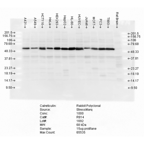
Western blot analysis of multiple cell lines lysates showing detection of Calreticulin protein using Rabbit Anti-Calreticulin Polyclonal Antibody (SPC-122). Load: 15 µgprotein. Block: 1.5% BSA for 30 minutes at RT. Primary Antibody: Rabbit Anti-Calreticulin Polyclonal Antibody (SPC-122) at 1:5000 for 2 hours at RT. Secondary Antibody: Donkey Anti-Rabbit IgG: HRP for 1 hour at RT.
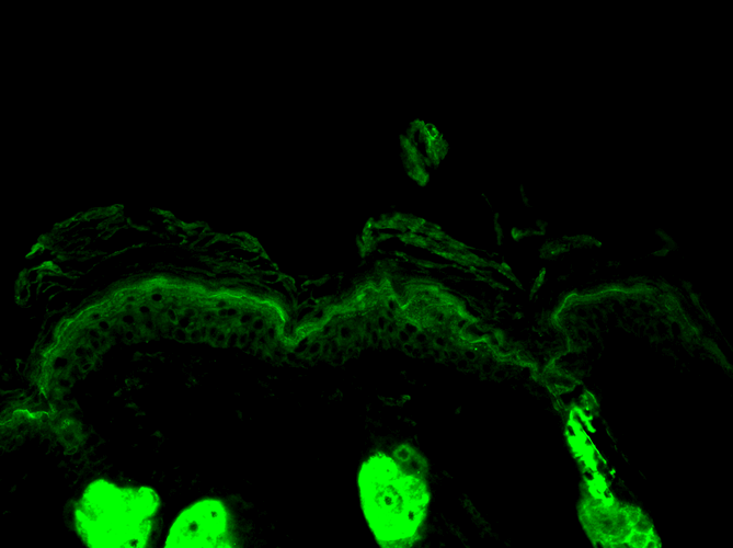
Immunohistochemistry analysis using Rabbit Anti-Calreticulin Polyclonal Antibody (SPC-122). Tissue: backskin. Species: Mouse. Fixation: Bouin’s Fixative Solution. Primary Antibody: Rabbit Anti-Calreticulin Polyclonal Antibody (SPC-122) at 1:100 for 1 hour at RT. Secondary Antibody: FITC Goat Anti-Rabbit (green) at 1:50 for 1 hour at RT. Localization: Cytoplasmic granule. Endoplasmic reticulum lumen.
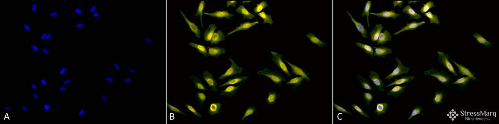
Immunocytochemistry/Immunofluorescence analysis using Rabbit Anti-Calreticulin Polyclonal Antibody (SPC-122). Tissue: Heat Shocked Cervical cancer cell line (HeLa). Species: Human. Fixation: 2% Formaldehyde for 20 min at RT. Primary Antibody: Rabbit Anti-Calreticulin Polyclonal Antibody (SPC-122) at 1:100 for 12 hours at 4°C. Secondary Antibody: R-PE Goat Anti-Rabbit (yellow) at 1:200 for 2 hours at RT. Counterstain: DAPI (blue) nuclear stain at 1:40000 for 2 hours at RT. Localization: Endoplasmic reticulum lumen. Cytoplasm. Magnification: 20x. (A) DAPI (blue) nuclear stain. (B) Anti-Calreticulin Antibody. (C) Composite. Heat Shocked at 42°C for 1h.
 Powered by Bioz
StressMarq Biosciences :
Based on validation through cited publications.