Discovery through Partnership | Excellence through Quality


Properties
| Storage Buffer | PBS pH7.4, 50% glycerol, 0.09% sodium azide *Storage buffer may change when conjugated |
| Storage Temperature | -20ºC, Conjugated antibodies should be stored according to the product label |
| Shipping Temperature | Blue Ice or 4ºC |
| Purification | Protein G Purified |
| Clonality | Monoclonal |
| Clone Number | N55/10 (Formerly sold as S55-10) |
| Isotype | IgG1 |
| Specificity | Detects ~260kDa. No cross-reactivity against Cav1.3. |
| Cite This Product | Cav3.2 Antibody (StressMarq Biosciences | Victoria, BC CANADA, Catalog# SMC-303, RRID: AB_2069540) |
| Certificate of Analysis | 1 µg/ml of SMC-303 was sufficient for detection of Cav3.2 in 10 µg of HEK cell lysate expressing Cav3.2 by colorimetric immunoblot analysis using Goat anti-mouse IgG:HRP as the secondary antibody. |
Biological Description
| Alternative Names | Voltage-dependent T-type calcium channel subunit alpha-1H, Voltage-gated calcium channel subunit alpha Cav3.2, CACNA1H, KIAA1120, Cav3.2, CACNA1HB, calcium channel, voltage-dependent, T type, alpha 1H subunit, alpha 1Hb subunit, ECA6, EIG6, FLJ90484, Low-voltage-activated calcium channel alpha1 3.2 subunit, low-voltage-activated calcium channel alpha13.2 subunit, voltage dependent t-type calcium channel alpha-1H subunit, voltage-dependent T-type calcium channel subunit alpha-1H, voltage-gated calcium channel alpha subunit Cav3.2, voltage-gated calcium channel alpha subunit CavT.2 |
| Research Areas | Calcium Channels, Cancer, Cell Signaling, Ion Channels, Neuroscience, Voltage-Gated Calcium Channels |
| Cellular Localization | Membrane |
| Accession Number | NP_001005407.1 |
| Gene ID | 8912 |
| Swiss Prot | O95180 |
| Scientific Background |
Cav3.2, encoded by the CACNA1H gene, is a low-voltage-activated T-type calcium channel that plays a critical role in regulating neuronal excitability, rhythmic firing, and calcium homeostasis. It is widely expressed in the brain and peripheral tissues, where it contributes to burst firing in thalamic neurons and modulates sensory signal transmission. Genetic studies have linked mutations in CACNA1H to childhood absence epilepsy, highlighting Cav3.2’s role in thalamocortical oscillations and seizure susceptibility. Beyond epilepsy, dysregulation of Cav3.2 expression and function has been associated with broader neurological and neurodegenerative conditions, including neuropathic pain, Parkinson’s disease, and Alzheimer’s disease. Its involvement in calcium dysregulation—a hallmark of neurodegeneration—positions Cav3.2 as a potential therapeutic target for modulating neuronal survival and excitability. Interestingly, Cav3.2 is also implicated in non-neuronal pathologies, such as prostate cancer progression, where its upregulation may support androgen-independent tumor growth. This underscores the channel’s broader physiological relevance and potential as a drug target across multiple disease domains. Despite its clinical significance, many aspects of Cav3.2’s in vivo function, regulatory mechanisms, and disease-specific roles remain under investigation. Advancing our understanding of Cav3.2 could open new avenues for targeted therapies in epilepsy, neurodegeneration, and beyond. |
| References |
1. Chen Y., et al. (2003) Ann. Neurol. 54(2): 239–43. 2. Khosravani H., et al. (2004) J Biol Chem. 279(11): 9681-9684. 3. Gackiere F., et al. (2008) J Biol Chem. 283(28): 19872. |
Product Images
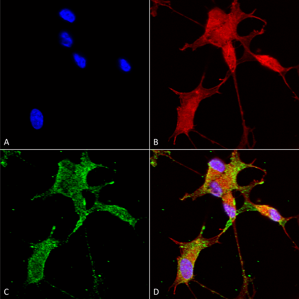
Immunocytochemistry/Immunofluorescence analysis using Mouse Anti-Cav3.2 Monoclonal Antibody, Clone N55/10 (SMC-303). Tissue: Neuroblastoma cells (SH-SY5Y). Species: Human. Fixation: 4% PFA for 15 min. Primary Antibody: Mouse Anti-Cav3.2 Monoclonal Antibody (SMC-303) at 1:50 for overnight at 4°C with slow rocking. Secondary Antibody: AlexaFluor 488 at 1:1000 for 1 hour at RT. Counterstain: Phalloidin-iFluor 647 (red) F-Actin stain; Hoechst (blue) nuclear stain at 1:800, 1.6mM for 20 min at RT. (A) Hoechst (blue) nuclear stain. (B) Phalloidin-iFluor 647 (red) F-Actin stain. (C) Cav3.2 Antibody (D) Composite.
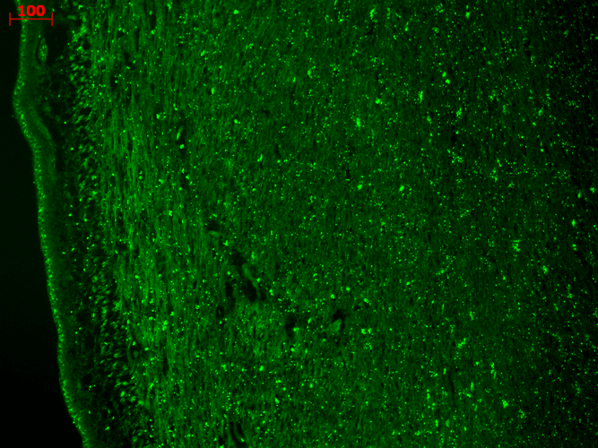
Immunohistochemistry analysis using Mouse Anti-CaV3.2 Calcium Channel Monoclonal Antibody, Clone N55/10 (SMC-303). Tissue: hippocampus. Species: Human. Fixation: Bouin’s Fixative and paraffin-embedded. Primary Antibody: Mouse Anti-CaV3.2 Calcium Channel Monoclonal Antibody (SMC-303) at 1:1000 for 1 hour at RT. Secondary Antibody: FITC Goat Anti-Mouse (green) at 1:50 for 1 hour at RT.
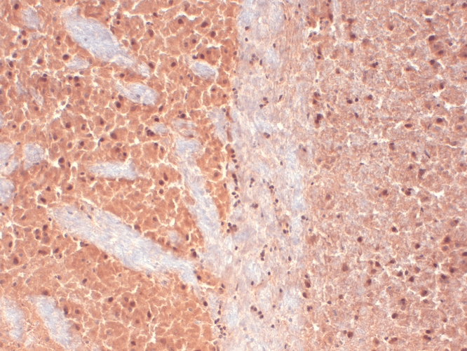
Immunohistochemistry analysis using Mouse Anti-CaV3.2 Calcium channel Monoclonal Antibody, Clone N55/10 (SMC-303). Tissue: frozen brain section. Species: Human. Fixation: 10% Formalin Solution for 12-24 hours at RT. Primary Antibody: Mouse Anti-CaV3.2 Calcium channel Monoclonal Antibody (SMC-303) at 1:1000 for 1 hour at RT. Secondary Antibody: HRP/DAB Detection System: Biotinylated Goat Anti-Mouse, Streptavidin Peroxidase, DAB Chromogen (brown) for 30 minutes at RT. Counterstain: Mayer Hematoxylin (purple/blue) nuclear stain at 250-500 µl for 5 minutes at RT.
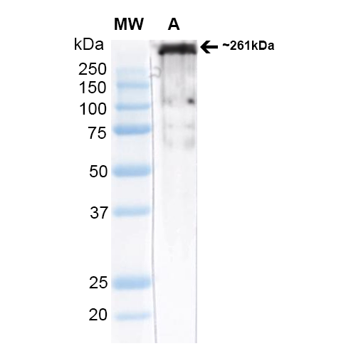
Western Blot analysis of Rat brain membrane lysate (native) showing detection of ~261 kDa Cav3.2 protein using Mouse Anti-Cav3.2 Monoclonal Antibody, Clone N55/10 (SMC-303). Block: 2% Skim Milk + 2% BSA in TBST. Primary Antibody: Mouse Anti-Cav3.2 Monoclonal Antibody (SMC-303) at 1:1000 for 2 hours at RT. Secondary Antibody: Anti-Mouse: HRP at 1:4000. Predicted/Observed Size: ~261 kDa.
 Powered by Bioz
StressMarq Biosciences :
Based on validation through cited publications.