Discovery through Partnership | Excellence through Quality


Properties
| Storage Buffer | PBS pH7.4, 50% glycerol, 0.09% sodium azide *Storage buffer may change when conjugated |
| Storage Temperature | -20ºC, Conjugated antibodies should be stored according to the product label |
| Shipping Temperature | Blue Ice or 4ºC |
| Purification | Protein G Purified |
| Clonality | Monoclonal |
| Clone Number | L62/29 (Formerly sold as S62-29) |
| Isotype | IgG1 |
| Specificity | Detects ~160kDa in rat brain membrane preparations. |
| Cite This Product | ATP7B Antibody (StressMarq Biosciences | Victoria, BC CANADA, Catalog# SMC-399, RRID: AB_11232610) |
| Certificate of Analysis | 1 µg/ml of SMC-399 was sufficient for detection of Copper-transporting ATPase2 in 20 µg of rat brain lysate by colorimetric immunoblot analysis using Goat IgG:HRP as the secondary antibody. |
Biological Description
| Alternative Names | ATP7B, ATPase Cu++ transporting beta polypeptide, ATPase Cu(2+) transporting beta polypeptide, Copper pump 2, Copper transporting ATPase 2, PWD, Toxic milk, tx, WC1, WD, Wilson disease associated protein, WND, WND/140 kDa |
| Research Areas | Cell Signaling, Ion Channels, Neuroscience |
| Cellular Localization | Cytoplasm, Golgi apparatus, Mitochondrion, Trans-golgi network membrane |
| Accession Number | NP_000044.2 |
| Gene ID | 540 |
| Swiss Prot | B7ZLR4 |
| Scientific Background |
ATP7B is a copper-transporting P-type ATPase critical for maintaining systemic and neuronal copper balance. Working in tandem with ATP7A, ATP7B facilitates the sequestration of intracellular copper into the vesicular secretory pathway, enabling its export and preventing toxic accumulation. This function is essential for copper detoxification, particularly in the liver, where ATP7B mediates biliary copper excretion. ATP7B operates by using ATP hydrolysis to transport copper ions across cellular membranes. Three isoforms of the ATP7B gene have been identified: isoform A is expressed in the liver, kidney, and brain; isoform B is brain-specific; and the WND/140 kDa isoform localizes to mitochondria, suggesting a role in mitochondrial copper regulation and oxidative stress response. Mutations in ATP7B cause Wilson disease, a rare autosomal recessive disorder characterized by copper accumulation in the liver and brain. Neurological manifestations include tremors, dystonia, psychiatric disturbances, and cognitive decline—highlighting the protein’s importance in neural function and copper detoxification. In neuroscience, ATP7B is increasingly recognized for its role in protecting neurons from copper-induced oxidative damage, a contributing factor in neurodegenerative diseases such as Alzheimer’s and Parkinson’s. Dysregulation of ATP7B may exacerbate mitochondrial dysfunction and protein aggregation, linking copper imbalance to broader neurodegenerative mechanisms. As a key regulator of copper metabolism, ATP7B represents a promising target for therapeutic strategies aimed at restoring metal homeostasis in neurodegenerative disease. |
| References |
1. Tanzi R.E., et al. (1993) Nature Genetics. 5: 344-350. 2. Ghr/nlm.gov/gene/ATP7B |
Product Images
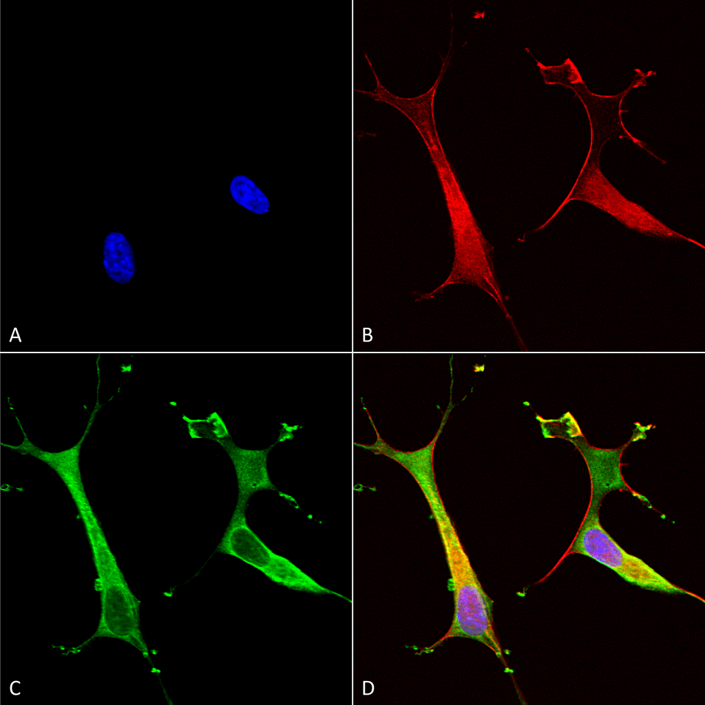
Immunocytochemistry/Immunofluorescence analysis using Mouse Anti-Copper Transporting ATPase 2 Monoclonal Antibody, Clone L62/29 (SMC-399). Tissue: Neuroblastoma cells (SH-SY5Y). Species: Human. Fixation: 4% PFA for 15 min. Primary Antibody: Mouse Anti-Copper Transporting ATPase 2 Monoclonal Antibody (SMC-399) at 1:100 for overnight at 4°C with slow rocking. Secondary Antibody: AlexaFluor 488 at 1:1000 for 1 hour at RT. Counterstain: Phalloidin-iFluor 647 (red) F-Actin stain; Hoechst (blue) nuclear stain at 1:800, 1.6mM for 20 min at RT. (A) Hoechst (blue) nuclear stain. (B) Phalloidin-iFluor 647 (red) F-Actin stain. (C) Copper Transporting ATPase 2 Antibody (D) Composite.
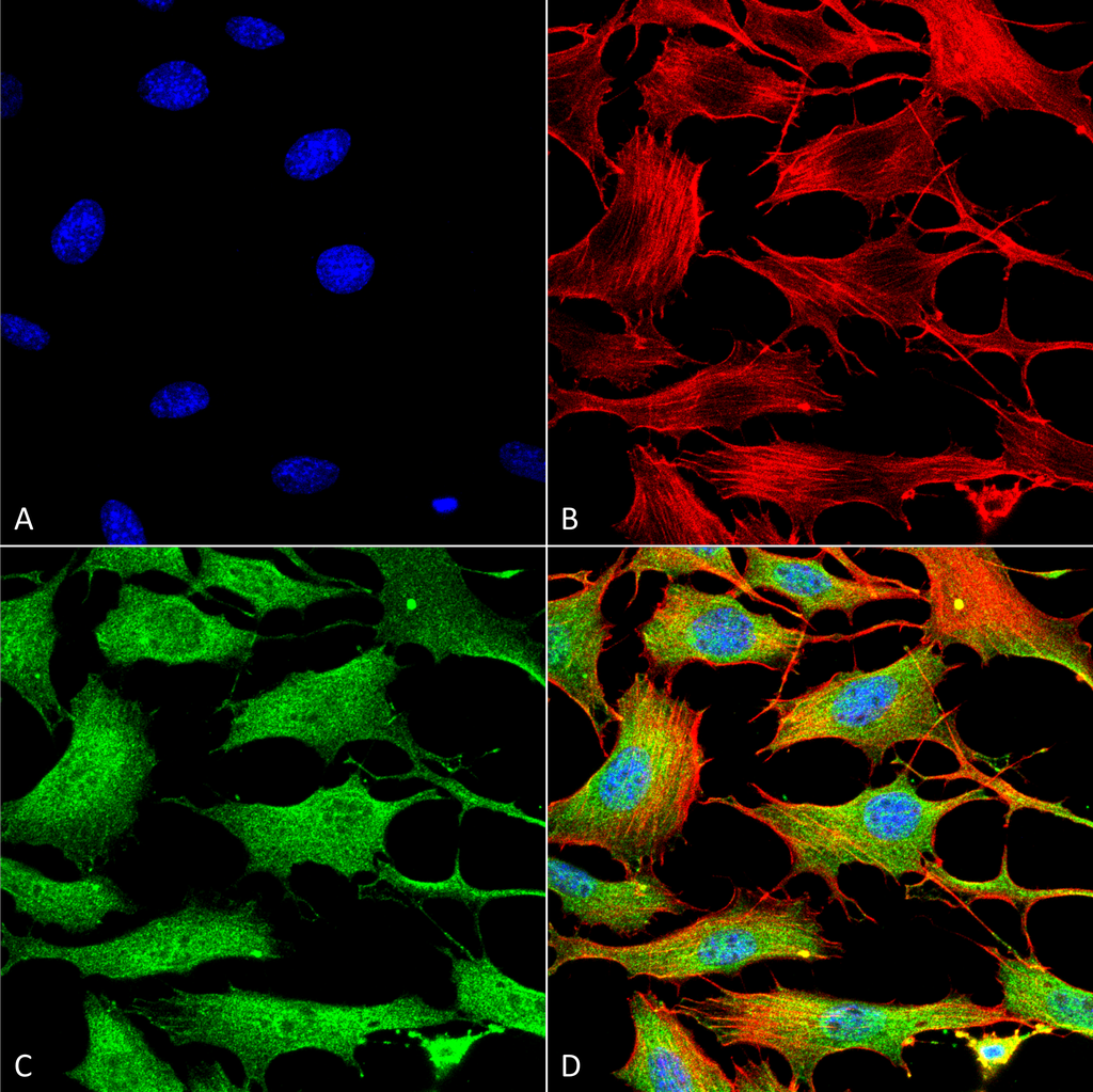
Immunocytochemistry/Immunofluorescence analysis using Mouse Anti-Copper Transporting ATPase 2 Monoclonal Antibody, Clone L62/29 (SMC-399). Tissue: NIH 3T3 (NIH 3T3). Species: Mouse. Fixation: 4% Formaldehyde for 15 min at RT. Primary Antibody: Mouse Anti-Copper Transporting ATPase 2 Monoclonal Antibody (SMC-399) at 1:100 for 60 min at RT. Secondary Antibody: Goat Anti-Mouse ATTO 488 at 1:200 for 60 min at RT. Counterstain: Phalloidin Texas Red F-Actin stain; DAPI (blue) nuclear stain at 1:1000, 1:5000 for 60 min at RT, 5 min at RT. Localization: Cytoplasm . Magnification: 60X. (A) DAPI (blue) nuclear stain. (B) Phalloidin Texas Red F-Actin stain. (C) Copper Transporting ATPase 2 Antibody. (D) Composite.
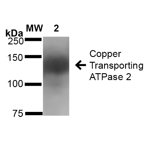
Western Blot analysis of Rat Brain Membrane showing detection of ~160 kDa Copper Transporting ATPase 2 protein using Mouse Anti-Copper Transporting ATPase 2 Monoclonal Antibody, Clone L62/29 (SMC-399). Lane 1: Molecular Weight Ladder (MW). Lane 2: Rat Brain Membrane cell lysate. Load: 20 µg. Block: 2% BSA and 2% Skim Milk in 1X TBST. Primary Antibody: Mouse Anti-Copper Transporting ATPase 2 Monoclonal Antibody (SMC-399) at 1:1000 for 16 hours at 4°C. Secondary Antibody: Goat Anti-Mouse IgG: HRP at 1:100 for 60 min at RT. Color Development: ECL solution for 6 min in RT. Predicted/Observed Size: ~160 kDa.
 Powered by Bioz
Reviews
There are no reviews yet.