Discovery through Partnership | Excellence through Quality


Properties
| Storage Buffer | PBS pH 7.4, 50% glycerol, 0.09% sodium azide *Storage buffer may change when conjugated |
| Storage Temperature | -20ºC, Conjugated antibodies should be stored according to the product label |
| Shipping Temperature | Blue Ice or 4ºC |
| Purification | Peptide Affinity Purified |
| Clonality | Polyclonal |
| Specificity | Binds to acetylated lysine 77 on HSP70. Detects 70 kDa band. |
| Cite This Product | HSP70 Antibody (Acetyl Lys77) (StressMarq Biosciences | Victoria, BC CANADA, Catalog# SPC-743, RRID: AB_2706545) |
| Certificate of Analysis | A 1:1000 dilution of SPC-743 was sufficient for detection of HSP70 Acetyl Lys77 in 10 µg of human HeLa cell lysates by ECL immunoblot analysis using goat anti-rabbit IgG:HRP as the secondary antibody. |
Biological Description
| Alternative Names | HSPA1A, HSPA1B, HSPA1, HSP70, HSP70-1, HSP70.1, HSP70-2, HSP72, HSP73, HSX70, Heat shock 70 kDa protein 1A, Heat shock 70 kDa protein 1B, HSP70 Acetylated lysine 77, HSP70 Acetyl Lys77 |
| Research Areas | Cancer, Cell Signaling, Chaperone Proteins, Heat Shock, Protein Trafficking, Tumor Biomarkers |
| Cellular Localization | Cytoplasm, Nucleus |
| Accession Number | NP_005336.3 |
| Gene ID | 3303 |
| Swiss Prot | P0DMV8/P0DMV9 |
| Scientific Background |
HSP70 proteins are a highly conserved family of 70-kDa molecular chaperones that play a central role in protein folding, stabilization, and transport across nearly all cellular compartments—including the cytosol, nucleus, mitochondria, endoplasmic reticulum, and chloroplasts. These proteins are encoded by a multigene family and are upregulated in response to cellular stress, making them key regulators of proteostasis. The HSP70 Acetylated Lysine 77 Antibody is a powerful tool for detecting HSP70 proteins specifically modified at lysine 77, a post-translational modification (PTM) that may influence chaperone activity, protein-protein interactions, and cellular localization. Acetylation at this site occurs within the highly conserved N-terminal ATP-binding domain, which is essential for HSP70’s conformational cycling and substrate release. This antibody may help researchers to monitor acetylation-specific regulation of HSP70 under stress conditions; investigate the role of lysine 77 acetylation in neurodegenerative diseases, cancer, and aging; or differentiate between modified and unmodified HSP70. |
| References |
1. Welch W.J. and Suhan J.P. (1986) J Cell Biol. 103: 2035-2050. 2. Boorstein W. R., Ziegelhoffer T. & Craig E. A. (1993) J.Mol. Evol. 38(1): 1-17. 3. Rothman J. (1989) Cell 59: 591-601. 4. DeLuca-Flaherty et al. (1990) Cell 62: 875-887. 5. Bork P., Sander C. & Valencia A. (1992) Proc. Nut1 Acad. Sci. USA 89: 7290-7294. 6. Fink A.L. (1999) Physiol. Rev. 79: 425-449. 7. Galan A., et al. (2000) J. Biol. Chem. 275: 11418-11424. 8. Kondo T., et al. (2000) J. Biol. Chem. 275: 8872-8879. 9. Misaki T., et al. (1994) Clin. Exp. Immun. 98: 234-239. 10. Pockley A.G., et al. (1998) Immunol. Invest. 27: 367-377. 11. Moon I.S., et al. (2001) Cereb Cortex 11(3): 238-248. 12. Dressel et al. (2000) J. Immunol. 164: 2362-2371. 13. Verma A.K., et al. (2007) Fish and Shellfish Immunology. 22(5): 547-555. 14. Banduseela V.C., et al. (2009) Physiol Genomics. 39(3): 141-159. |
Product Images
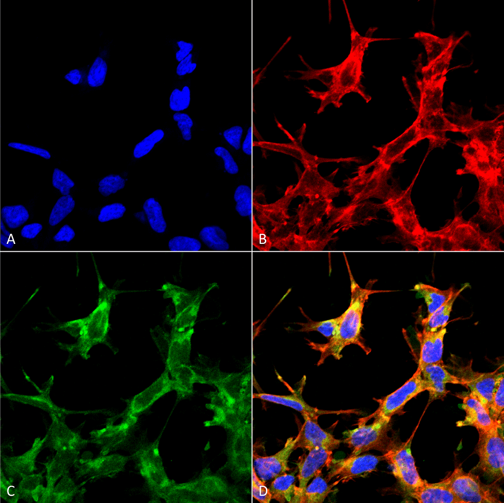
Immunocytochemistry/Immunofluorescence analysis using Rabbit Anti-HSP70 Acetyl Lys77 Polyclonal Antibody (SPC-743). Tissue: Embryonic kidney cells (HEK293) cultured overnight with 50 µM H2O2. Species: Human. Fixation: 5% Formaldehyde for 5 min. Primary Antibody: Rabbit Anti-HSP70 Acetyl Lys77 Polyclonal Antibody (SPC-743) at 1:60 for 30-60 min at RT. Secondary Antibody: Goat Anti-Rabbit Alexa Fluor 488 at 1:1500 for 30-60 min at RT. Counterstain: Phalloidin Alexa Fluor 633 F-Actin stain; DAPI (blue) nuclear stain at 1:250, 1:50000 for 30-60 min at RT. Localization: Cytoplasmic. Magnification: 20X (2X Zoom). (A) DAPI (blue) nuclear stain. (B) Phalloidin Alex Fluor 633 F-Actin stain. (C) HSP70 Antibody (Acetyl Lys77). (D) Composite. Courtesy of: Dr. Robert Burke, University of Victoria.
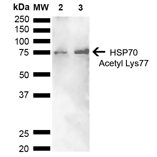
Western blot analysis of Human Cervical cancer cell line (HeLa) lysate showing detection of ~70 kDa HSP70 Acetyl Lys77 protein using Rabbit Anti-HSP70 Acetyl Lys77 Polyclonal Antibody (SPC-743). Lane 1: Molecular Weight Ladder (MW). Lane 2: Cervical Cancer cell line (HeLa) lysate. Lane 3: H2O2 Cervical Cancer cell line (HeLa) lysate. Load: 10 µg. Block: 5% Skim Milk in 1X TBST. Primary Antibody: Rabbit Anti-HSP70 Acetyl Lys77 Polyclonal Antibody (SPC-743) at 1:1000 for 2 hours at RT. Secondary Antibody: Goat Anti-Rabbit HRP:IgG at 1:3000 for 1 hour at RT. Color Development: ECL solution for 5 min at RT. Predicted/Observed Size: ~70 kDa.
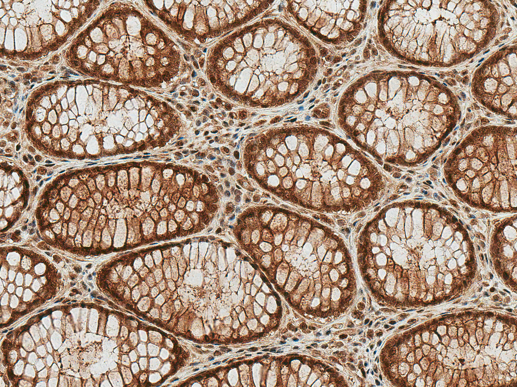
Immunohistochemistry analysis using Rabbit Anti-HSP70 (Acetyl Lys77) Polyclonal Antibody (SPC-743). Tissue: Colon Cancer. Species: Human. Fixation: Formalin Fixed Paraffin-Embedded. Primary Antibody: Rabbit Anti-HSP70 (Acetyl Lys77) Polyclonal Antibody (SPC-743) at 1:50 for 30 min at RT. Counterstain: Hematoxylin. Magnification: 20X. HRP-DAB Detection.
 Powered by Bioz
StressMarq Biosciences :
Based on validation through cited publications.