Discovery through Partnership | Excellence through Quality


Properties
| Storage Buffer | PBS pH7.2, 50% glycerol, 0.09% sodium azide *Storage buffer may change when conjugated |
| Storage Temperature | -20ºC, Conjugated antibodies should be stored according to the product label |
| Shipping Temperature | Blue Ice or 4ºC |
| Purification | Protein G Purified |
| Clonality | Monoclonal |
| Clone Number | Hyb-K41220A |
| Isotype | IgG2a |
| Specificity | Detects 90kDa. Will detect both alpha (inducible) and beta (constitutively-expressed) forms. |
| Cite This Product | HSP90 alpha/beta Antibody (StressMarq Biosciences | Victoria, BC CANADA, Catalog# SMC-135, RRID: AB_2121063) |
| Certificate of Analysis | 1 µg/ml was sufficient for detection of HSP90αβ in 20 µg of heat shocked HeLa cell lysate by colorimetric immunoblot analysis using Goat Anti-Mouse IgG:HRP as the secondary. |
Biological Description
| Alternative Names | HSP90AA1, HSP90AB1, HSP90-alpha, HSP90-beta, HSP90A, HSP90B, HSP89A, HSP86, HSP84, HSP90Alpha, HSPCA, HSPC1, HSPC2, HSPCB, HSPCAL3, Heat shock protein HSP 90-alpha, Heat shock protein HSP 90-beta, Heat shock 90 kDa protein 1 alpha isoform, Heat shock 84 kDa protein, D6S182, FLJ26984 |
| Research Areas | Cancer, Cell Signaling, Chaperone Proteins, Heat Shock, Protein Trafficking, Tumor Biomarkers |
| Cellular Localization | Cytoplasm, Melanosome |
| Accession Number | NP_031381.2, NP_001017963.2 |
| Gene ID | 3326, 3320 |
| Swiss Prot | P08238, P07900 |
| Scientific Background |
HSP90α and HSP90β are the two major cytosolic isoforms of the highly conserved heat shock protein 90 (HSP90) family, essential for maintaining protein homeostasis in all eukaryotic cells. While they share 85% amino acid sequence identity, the isoforms differ in structure and function: HSP90α primarily forms homodimers and is stress-inducible, whereas HSP90β exists mainly as a monomer and is constitutively expressed. Both isoforms are abundantly expressed in the brain and play critical roles in the folding, maturation, and stabilization of a wide range of client proteins involved in neuronal signaling, synaptic plasticity, and cellular stress responses. These include kinases (e.g., c-Raf), transcription factors (e.g., p53), and hormone receptors—many of which are implicated in neurodegenerative diseases such as Alzheimer’s, Parkinson’s, and Huntington’s disease. HSP90α and HSP90β function as part of dynamic chaperone complexes, interacting with co-chaperones like Cdc37 and p23. ATP binding drives conformational changes that enable these complexes to stabilize client proteins and prevent their degradation. However, in disease contexts, this protective mechanism can inadvertently preserve toxic protein species, contributing to pathogenesis. By recognizing both HSP90α and HSP90β, this antibody provides a powerful tool for researchers exploring the dual roles of HSP90 in neuronal health and disease. |
| References |
1. Nemoto, T. et al. (1997) J.Biol Chem. 272: 26179-26187. 2. Minami Y, et al. (1991), J.Biol Chem. 266: 10099-10103. 3. Arlander SJH, et al. (2003) J Biol Chem 278: 52572-52577. 4. Pearl H, et al. (2001) Adv Protein Chem 59: 157-186. 5. Neckers L, et al. (2002) Trends Mol Med 8: S55-S61. 6. Pratt W, Toft D. (2003) Exp Biol Med 228: 111-133. 7. Pratt W, Toft D. (1997) Endocr Rev 18: 306–360. 8. Pratt WB. (1998) Proc Soc Exptl Biol Med 217: 420–434. 9. Whitesell L, et al. (1994) Proc Natl Acad Sci USA 91: 8324–8328. 10. Kishimoto J, et al. (2005). Cell Stress and Chaperones. 10 (4): 296-311. |
Product Images
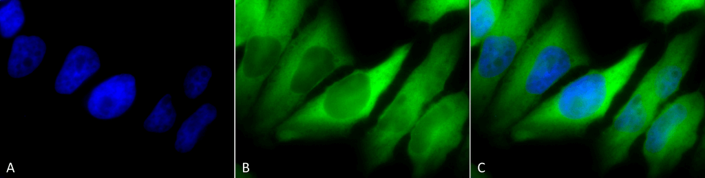
Immunocytochemistry/Immunofluorescence analysis using Mouse Anti-Hsp90 alpha/beta Monoclonal Antibody, Clone K41220A (SMC-135). Tissue: Cervical cancer cell line (HeLa). Species: Human. Fixation: 2% Formaldehyde for 20 min at RT. Primary Antibody: Mouse Anti-Hsp90 alpha/beta Monoclonal Antibody (SMC-135) at 1:100 for 12 hours at 4°C. Secondary Antibody: FITC Goat Anti-Mouse (green) at 1:200 for 2 hours at RT. Counterstain: DAPI (blue) nuclear stain at 1:40000 for 2 hours at RT. Localization: Cytoplasm. Melanosome. Magnification: 100x. (A) DAPI (blue) nuclear stain. (B) Anti-Hsp90 alpha/beta Antibody. (C) Composite.
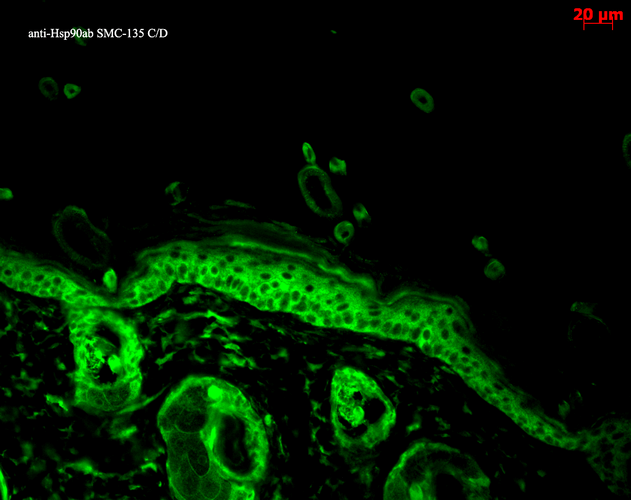
Immunohistochemistry analysis using Mouse Anti-Hsp90 alpha Monoclonal Antibody, Clone K41220A (SMC-135). Tissue: backskin. Species: Mouse. Fixation: Bouin’s Fixative and paraffin-embedded. Primary Antibody: Mouse Anti-Hsp90 alpha Monoclonal Antibody (SMC-135) at 1:100 for 1 hour at RT. Secondary Antibody: FITC Goat Anti-Mouse (green) at 1:50 for 1 hour at RT. Localization: Epidermis.
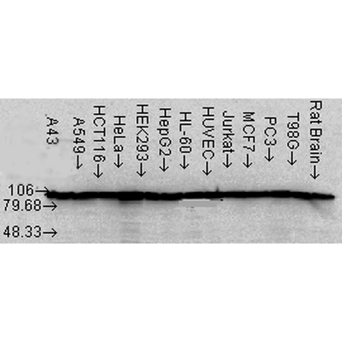
Western Blot analysis of Human Cell lysates showing detection of Hsp90 alpha protein using Mouse Anti-Hsp90 alpha Monoclonal Antibody, Clone K41220A (SMC-135). Load: 15 µg. Block: 1.5% BSA for 30 minutes at RT. Primary Antibody: Mouse Anti-Hsp90 alpha Monoclonal Antibody (SMC-135) at 1:1000 for 2 hours at RT. Secondary Antibody: Sheep Anti-Mouse IgG: HRP for 1 hour at RT.
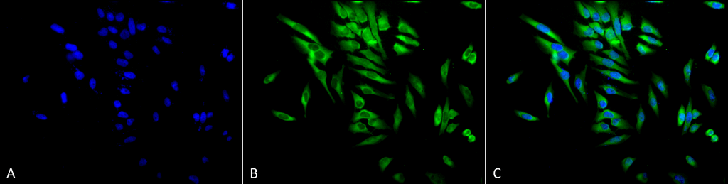
Immunocytochemistry/Immunofluorescence analysis using Mouse Anti-Hsp90 alpha/beta Monoclonal Antibody, Clone K41220A (SMC-135). Tissue: Cervical cancer cell line (HeLa). Species: Human. Fixation: 2% Formaldehyde for 20 min at RT. Primary Antibody: Mouse Anti-Hsp90 alpha/beta Monoclonal Antibody (SMC-135) at 1:100 for 12 hours at 4°C. Secondary Antibody: FITC Goat Anti-Mouse (green) at 1:200 for 2 hours at RT. Counterstain: DAPI (blue) nuclear stain at 1:40000 for 2 hours at RT. Localization: Cytoplasm. Melanosome. Magnification: 20x. (A) DAPI (blue) nuclear stain. (B) Anti-Hsp90 alpha/beta Antibody. (C) Composite.
 Powered by Bioz
StressMarq Biosciences :
Based on validation through cited publications.