Discovery through Partnership | Excellence through Quality


Properties
| Storage Buffer | PBS pH 7.4, 50% glycerol, 0.09% Sodium azide *Storage buffer may change when conjugated |
| Storage Temperature | -20ºC, Conjugated antibodies should be stored according to the product label |
| Shipping Temperature | Blue Ice or 4ºC |
| Purification | Affinity Purified |
| Clonality | Recombinant Monoclonal |
| Clone Number | J18 |
| Isotype | IgG |
| Specificity | Binds to phosphorylated serine 129 on alpha synuclein. Does not detect unphosphorylated serine 129 alpha synuclein |
| Cite This Product | Alpha Synuclein Antibody (pSer129) (StressMarq Biosciences | Victoria, BC CANADA, Catalog# SMC-600, RRID: AB_2820299) |
| Certificate of Analysis | A 1:500 dilution of SMC-600 was sufficient for detection of Alpha Synuclein pSer129 in 10 µg of Mouse Brain by ECL immunoblot analysis using Goat Anti-Rabbit IgG:HRP as the secondary antibody. |
Biological Description
| Alternative Names | Alpha Synuclein, α-Synuclein, SNCA, alphaSYN, NACP, Non-A beta component of AD amyloid, Non-A4 component of amyloid precursor, Phosphorylated alpha synuclein, Phospho-alpha Synuclein (S129), Alpha-synuclein (phospho S129), Alpha Synuclein (phospho Ser129), Alpha-Synuclein Phospho Ser129, phospho-α-Synuclein (Ser129), Alpha Synuclein phospho Serine 129, Alpha Synuclein phospho Ser 129, Alpha Synuclein pSerine 129, Alpha Synuclein pSer 129, Alpha Synuclein phosphoSer 129, isoform NACP140, PARK1, PARK 1, PARK4, PARK 4, Parkinson disease (autosomal dominant, Lewy body) 4, Parkinson disease familial 1, SYN, Synuclein alpha, Synuclein alpha 140, Synuclein, alpha (non A4 component of amyloid precursor), SYUA_HUMAN |
| Research Areas | Alzheimer's Disease, Neurodegeneration, Neuroscience, Parkinson's Disease, Synuclein, Tangles & Tau, Multiple System Atrophy |
| Cellular Localization | Cell Junction, Cytoplasm, Cytosol, Membrane, Nucleus, Synapse |
| Accession Number | NP_000336.1 |
| Gene ID | 6622 |
| Swiss Prot | P37840 |
| Scientific Background |
Alpha-synuclein (SNCA) is a neuronal protein predominantly expressed in the brain, where it is enriched at presynaptic terminals and plays a key role in synaptic function (1). It is also highly expressed in mitochondria-rich regions such as the olfactory bulb, hippocampus, striatum, and thalamus, suggesting a role in mitochondrial dynamics and neuronal energy metabolism (2). Functionally, alpha-synuclein interacts with tubulin (3), indicating a potential role as a microtubule-associated protein involved in cytoskeletal organization and intracellular transport. It is essential for cognitive development, with SNCA inactivation linked to impaired spatial learning and working memory (4). Pathologically, SNCA aggregates are a major non-Aβ component of amyloid plaques in Alzheimer’s disease and a defining feature of Lewy body inclusions in Parkinson’s disease (PD). PD is characterized by the progressive accumulation of alpha-synuclein and ubiquitin-positive inclusions in vulnerable neurons, contributing to neurodegeneration (5, 6). Critically, phosphorylation of alpha-synuclein at serine 129 (pSer129) is a key post-translational modification associated with disease pathology. Approximately 90% of alpha-synuclein found in Lewy bodies is phosphorylated at Ser129, compared to only a small fraction in the healthy brain (7, 8). This modification is widely used as a biomarker for pathological alpha-synuclein and is a focal point in therapeutic and diagnostic research targeting synucleinopathies. |
| References |
1. “Genetics Home Reference: SNCA”. US National Library of Medicine. (2013). 2. Zhang L., et al. (2008) Brain Res. 1244: 40-52. 3. Alim M.A., et al. (2002) J Biol Chem. 277(3): 2112-2117. 4. Kokhan V.S., Afanasyeva M.A., Van’kin G. (2012) Behav. Brain. Res. 231(1): 226-230. 5. Spillantini M.G., et al. (1997) Nature. 388(6645): 839-840. 6. Mezey E., et al. (1998) Nat Med. 4(7): 755-757. 7. Fujiwara H., et al. (2002) Nat Cell Biol. 4(2):160-4. 8. Anderson J.P., et al. (2006) J Biol Chem. 281(40):29739-52. |
Product Images
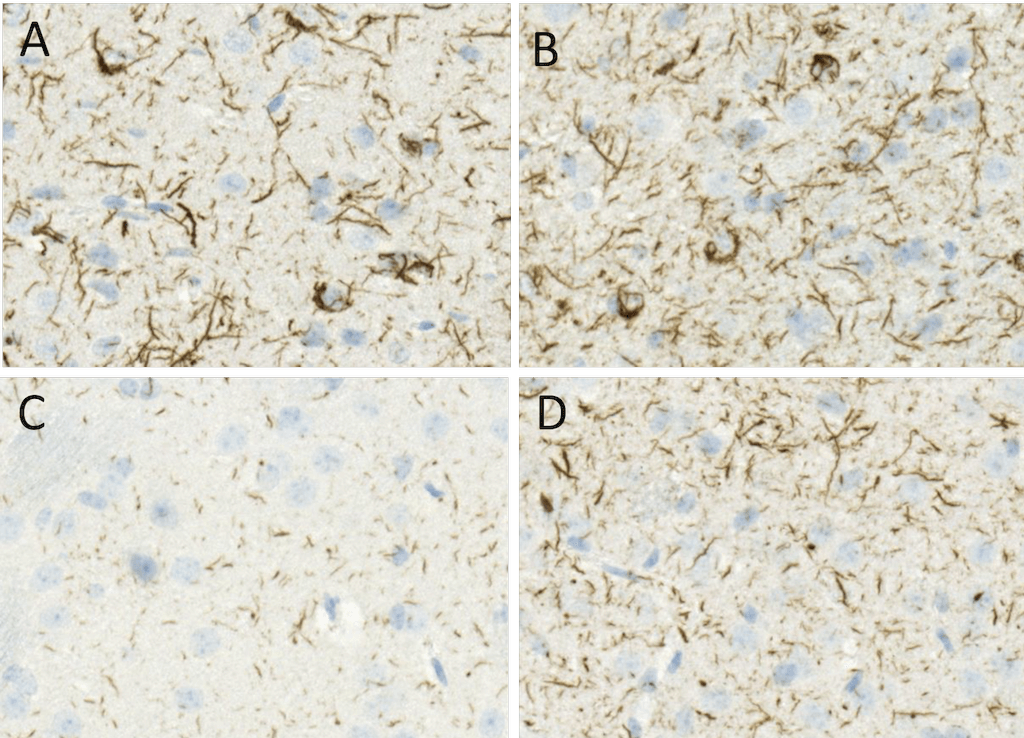
Immunohistochemistry analysis using Rabbit Anti-Alpha Synuclein pSer129 Monoclonal Antibody, Clone J18 (SMC-600). Tissue: Brain. Species: Mouse. Primary Antibody: Rabbit Anti-Alpha Synuclein pSer129 Monoclonal Antibody (SMC-600) at 1:10000. Secondary Antibody: anti-rabbit HRP. C57/BL6 mice were injected with 5 ug sonicated mouse recombinant alpha synuclein PFFs (SPR-324) at 8 weeks of age. Mice were unilaterally injected in the dorsal striatum (bregma AP + 0.2 mm, L +/1 2.0 mm, V – 3.0 mm) and sacrificed 30 days post-injection. (A) contralateral cortex. (B) ipsilateral cortex. (C) contralateral striatum. (D) ipsilateral striatum. Courtesy of: Porsolt.
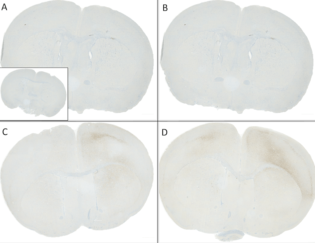
Immunohistochemistry analysis using Rabbit Anti-Alpha Synuclein pSer129 Monoclonal Antibody, Clone J18 (SMC-600). Tissue: Brain. Species: Mouse. Primary Antibody: Rabbit Anti-Alpha Synuclein pSer129 Monoclonal Antibody (SMC-600) at 1:10000. Secondary Antibody: anti-rabbit HRP. C57/BL6 mice were injected with sonicated recombinant mouse alpha synuclein monomers or fibrils at 8 weeks of age. Mice were unilaterally injected in the dorsal striatum (bregma AP + 0.2 mm, L +/1 2.0 mm, V – 3.0 mm) and sacrificed 30 days post-injection. (A) 1.25 uL mouse alpha synuclein monomers (SPR-323). (B) 2.5 uL mouse alpha synuclein monomers (SPR-323). (C) 2.5 ug alpha synuclein PFFs (SPR-324). (D) 5 ug alpha synuclein PFFs (SPR-324). Inset: PBS (negative control). Courtesy of: Porsolt.
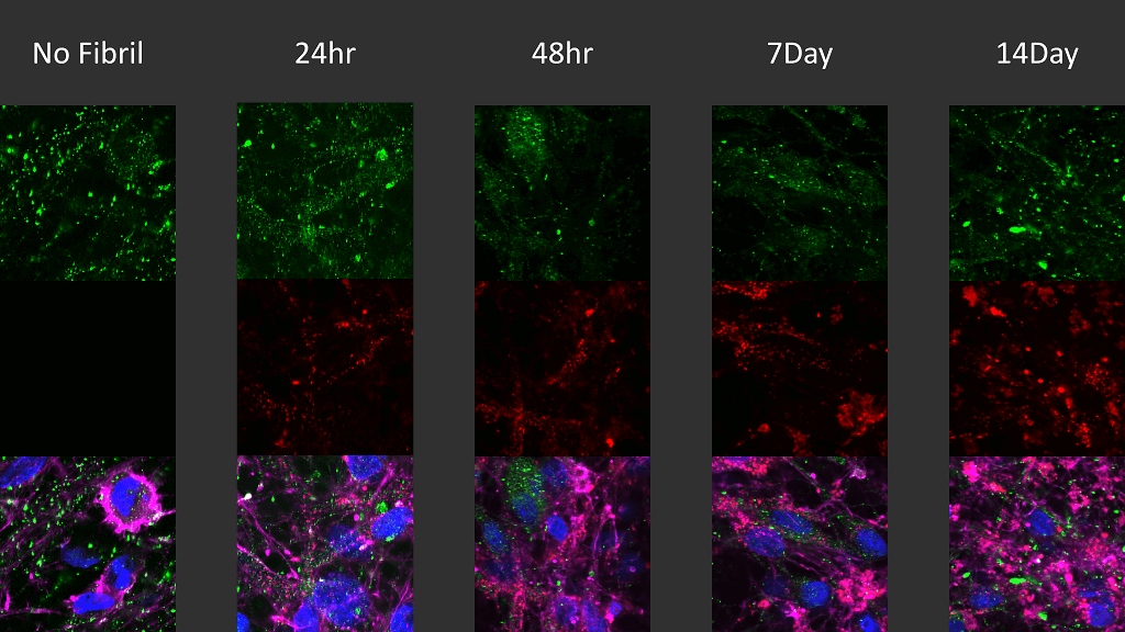
Immunocytochemistry / Immunofluorescence analysis of human iPSC-derived neurons (Stem Cell Catalog Code ASE-9321KF) treated with 2.5µg ATTO 594 labeled type I alpha-synuclein pre-formed fibrils (SPR-322-A594) for up to 14 days. Cells seeded at 8k cells per well. Green: mouse anti-alpha synuclein (pSer129) monoclonal antibody (SMC-600) 1:5000; Red: alpha-synuclein PFFs (SPR-322-A594); Pink: actin; Blue: Hoechst / DNA.
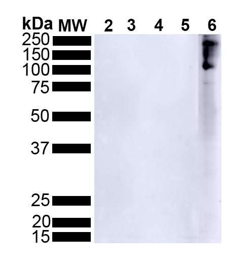
Western Blot analysis of Human Alpha Synuclein showing detection of Alpha Synuclein pSer129 protein using Rabbit Anti-Alpha Synuclein pSer129 Monoclonal Antibody, Clone J18 (SMC-600). Lane 1: MW ladder. Lane 2: 0.5 ug human alpha synuclein monomer (SPR-321). Lane 3: 2 ug human alpha synuclein monomer (SPR-321). Lane 4: 0.5 ug human alpha synuclein PFFs (SPR-322). Lane 5: 2 ug human alpha synuclein PFFs (SPR-322). Lane 6: 15 ug human Parkinson’s Disease brain lysate.. Block: 5% BSA in TBST. Primary Antibody: Rabbit Anti-Alpha Synuclein pSer129 Monoclonal Antibody (SMC-600) at 1:500 for 2 hours at RT with shaking. Secondary Antibody: Goat anti-rabbit IgG:HRP at 1:4000 for 1 hour at RT with shaking. Color Development: Chemiluminescent for HRP (Moss) for 5 min in RT.
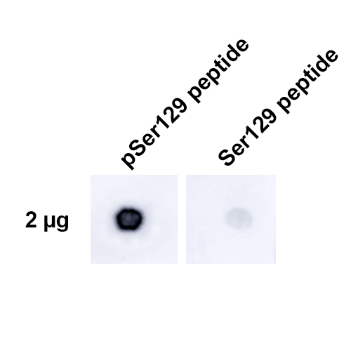
Dot Blot analysis using Rabbit Anti-Alpha Synuclein pSer129 Monoclonal Antibody, Clone J18 (SMC-600). Tissue: alpha synuclein peptide. Primary Antibody: Rabbit Anti-Alpha Synuclein pSer129 Monoclonal Antibody (SMC-600) at 1:500 for 2 hours at RT with shaking . Secondary Antibody: Goat anti-rabbit IgG:HRP at 1:4000 for 1 hour at RT with shaking . Phospho peptide sequence: AYEMP-pS-EEGYQ. Non-phospho peptide sequence: AYEMPSEEGYQ. This sequence is the same for human, mouse, and rat.
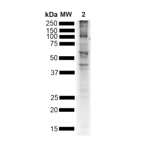
Western Blot analysis of Mouse Brain showing detection of Alpha Synuclein pSer129 protein using Rabbit Anti-Alpha Synuclein pSer129 Monoclonal Antibody, Clone J18 (SMC-600). Lane 1: MW ladder. Lane 2: Mouse brain. Load: 15 ug. Block: 5% BSA in TBST. Primary Antibody: Rabbit Anti-Alpha Synuclein pSer129 Monoclonal Antibody (SMC-600) at 1:500 for 2 hours at RT with shaking. Secondary Antibody: Goat anti-rabbit IgG:HRP at 1:4000 for 1 hour at RT with shaking. Color Development: Chemiluminescent for HRP (Moss) for 5 min in RT.
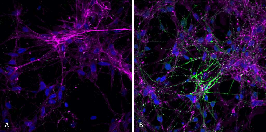
Immunocytochemistry/Immunofluorescence analysis using Rabbit Anti-Alpha Synuclein (pSer129) Monoclonal Antibody, Clone J18 (SMC-600). Tissue: iPSC-derived neurons. Species: Human. Primary Antibody: Rabbit Anti-Alpha Synuclein (pSer129) Monoclonal Antibody (SMC-600) at 1:1000 for O/N at 4°C. Secondary Antibody: Anti-Rabbit: A488 at 1:1000 for 1 hour at RT. Magnification: 40X. Nuclear stain: Hoechst- 20 min, RT (blue). Actin stain: Phalloidin-647- 20 min, RT (magenta). 4K cells per well. iPSC neurons: Applied StemCell Catalog # ASE-9321K. A) negative control; no fibrils added to well. B) 7 days after addition of active recombinant human pre-formed fibrils (Type 1). StressMarq catalog # SPR-322. Sonicated before use, 2.5 ug per well.
 Powered by Bioz
StressMarq Biosciences :
Based on validation through cited publications.