Discovery through Partnership | Excellence through Quality


Properties
| Storage Buffer | PBS pH7.4, 50% glycerol, 0.09% sodium azide *Storage buffer may change when conjugated |
| Storage Temperature | -20ºC, Conjugated antibodies should be stored according to the product label |
| Shipping Temperature | Blue Ice or 4ºC |
| Purification | Protein G Purified |
| Clonality | Monoclonal |
| Clone Number | 6G9 |
| Isotype | IgG1 |
| Specificity | Detects ~50-60kDa. Recognizes both phosphorylated and non-phosphorylated forms. |
| Cite This Product | CaMKII alpha Antibody (StressMarq Biosciences | Victoria, BC CANADA, Catalog# SMC-124, RRID: AB_2275062) |
| Certificate of Analysis | 0.1 µg/ml was sufficient for detection of CamKII in 20 µg rat brain tissue extract by colorimetric immunoblot analysis using Goat Anti-Mouse IgG:AP as the secondary. |
Biological Description
| Alternative Names | CaMKII, CaM kinase II, CaM KII, CaMK-II, CamK-II, CAMKII, CaMKii, CamKII, Camkii, camKII, camkII, camkii, CAMKIIalpha, CamKIIalpha, CAMKIId, CDPK1, dCAMKII, dCaMKII, DCK, CAMK2A, CAMK2B, CAMK2D, CAMK2G, CAMKA |
| Research Areas | Cell Signaling, Phosphorylation, Post-translational Modifications |
| Cellular Localization | Cell Junction, Cytoplasm, Mitochondrion, Nucleus, Presynaptic Cell Membrane, Synapse |
| Accession Number | NP_033922.1 |
| Gene ID | 12322 |
| Swiss Prot | P11798 |
| Scientific Background |
Calcium/calmodulin-dependent protein kinase II (CaMKII) is a pivotal serine/threonine kinase that plays a critical role in synaptic plasticity, memory formation, and neurodegenerative disease mechanisms. Highly enriched in forebrain neurons, CaMKII is localized to both the soma and dendrites, where it orchestrates calcium signaling in response to synaptic activity. The neuronal isoforms—comprising 52 kDa (α subunit) and 60 kDa (β subunit)—assemble into multimeric holoenzymes that integrate calcium dynamics with downstream signaling. CaMKII activation is initiated by Ca²⁺/calmodulin binding, which relieves autoinhibition and triggers autophosphorylation at threonine 286 (Thr286). This phosphorylation event confers autonomous kinase activity, even after calcium levels subside, and enhances CaMKII’s affinity for NMDA receptors at postsynaptic densities—an essential mechanism for long-term potentiation (LTP). The phosphorylation state of Thr286 is tightly regulated: protein phosphatase 1 (PP1) dephosphorylates it, while protein kinase A (PKA) counteracts this dephosphorylation, sustaining CaMKII activity. Dysregulation of CaMKII signaling has been implicated in multiple neurodegenerative disorders, including Alzheimer’s disease, where aberrant phosphorylation and synaptic dysfunction are hallmark features. As a master regulator of synaptic strength and plasticity, CaMKII remains a high-value target in neuroscience research and therapeutic development for cognitive and neurodegenerative disorders. |
| References |
1. Hughes K. et al. (2001) J. Biol. Chem. 276: 36008–36013. 2. Barria A. et al. (1997) Science 276: 2042–2045. 3. Bennet M.K. and Kennedy M.B. (1987) Proc. Natl. Acad. Sci. U.S.A. 84: 1794-1798. 4. Broke L., Srinivasan M. and Schulman H. (1995) J. Neurosci. 15: 6797-6808. 5. Nghiem P., Saati S. M., Martens C. L., Gardner P. and Schulman H. (1993) J. Biol. Chem. 268: 5471-5479. 6. Edman C.F. and Schulman H. (1994) Biochem. Biophys. Acta 1221: 90-102. 7. Tombes R.M. and Krystal G.W., (1997) Biochem. Biophys. Acta 13555: 281-292. 8. Means A.R. (2000) Mol. Endocrinol. 14: 4–12. 9. Makhinson M. et al. (1999) J. Neurosci. 19: 2500–2510. 10. Strack S. and Colbran R.J. (1998) J. Biol. Chem. 273: 20689-20692. 11. Leonard S.A., Lim I.A., Hemsworth D.E., Horne M.C. and Hell J.W. (1999) Proc. Natl. Acad. Sci. U.S.A. 96: 3239-3244. 12. Shen K. and Meyer Y. (1999) Science 284: 162-167. |
Product Images
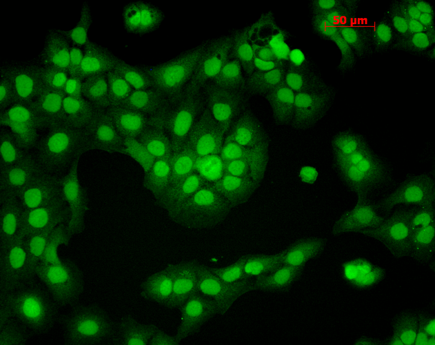
Immunocytochemistry/Immunofluorescence analysis using Mouse Anti-CaMKII Monoclonal Antibody, Clone 6G9 (SMC-124). Tissue: HaCaT cells. Species: Human. Fixation: Cold 100% methanol for 10 minutes at -20°C. Primary Antibody: Mouse Anti-CaMKII Monoclonal Antibody (SMC-124) at 1:100 for 1 hour at RT. Secondary Antibody: FITC Goat Anti-Mouse (green) at 1:50 for 1 hour at RT. Localization: Nuclear Staining.
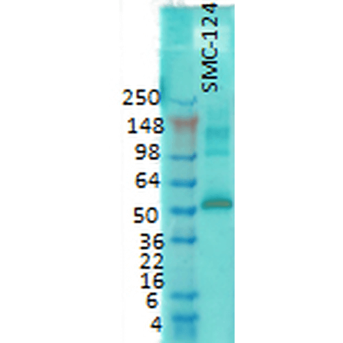
Western Blot analysis of Rat brain membrane lysate showing detection of CaMKII protein using Mouse Anti-CaMKII Monoclonal Antibody, Clone 6G9 (SMC-124). Primary Antibody: Mouse Anti-CaMKII Monoclonal Antibody (SMC-124) at 1:1000.
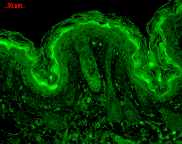
Immunohistochemistry analysis using Mouse Anti-CaMKII Monoclonal Antibody, Clone 6G9 (SMC-124). Tissue: backskin. Species: Mouse. Fixation: Bouin’s Fixative and paraffin-embedded. Primary Antibody: Mouse Anti-CaMKII Monoclonal Antibody (SMC-124) at 1:100 for 1 hour at RT. Secondary Antibody: FITC Goat Anti-Mouse (green) at 1:50 for 1 hour at RT. Localization: Hair follicles, epidermis.
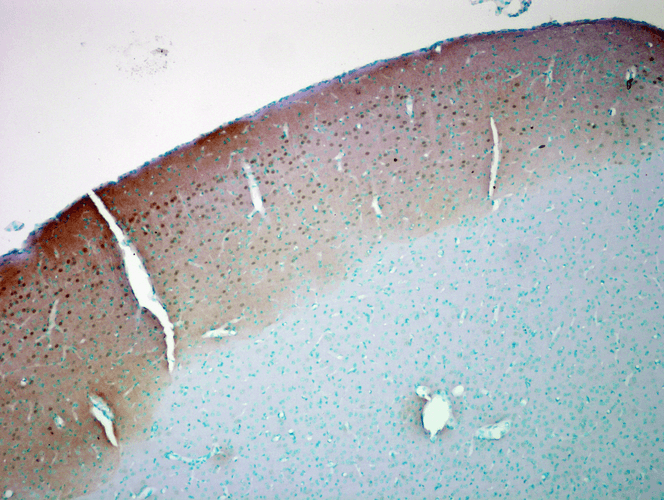
Immunohistochemistry analysis using Mouse Anti-CaMKII Monoclonal Antibody, Clone 6G9 (SMC-124). Tissue: colon carcinoma. Species: Human. Fixation: Formalin. Primary Antibody: Mouse Anti-CaMKII Monoclonal Antibody (SMC-124) at 1:10000 for 12 hours at 4°C. Secondary Antibody: Biotin Goat Anti-Mouse at 1:2000 for 1 hour at RT. Counterstain: Mayer Hematoxylin (purple/blue) nuclear stain at 200 µl for 2 minutes at RT. Magnification: 40x.
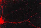
Immunocytochemistry/Immunofluorescence analysis using Mouse Anti-CaMKII Monoclonal Antibody, Clone 6G9 (SMC-124). Tissue: dissociated hippocampal neurons. Species: Mouse. Fixation: Cold 4% paraformaldehyde/0.2% glutaraldehyde in 0.1M sodium phosphate buffer. Primary Antibody: Mouse Anti-CaMKII Monoclonal Antibody (SMC-124) at 1:1000 for 12 hours at 4°C. Secondary Antibody: FITC Goat Anti-Mouse IgG (green) at 1:50 for 30 minutes at RT. Magnification: 10X. Courtesy of: Mary Kennedy, Caltech.
 Powered by Bioz
StressMarq Biosciences :
Based on validation through cited publications.