Properties
| Storage Buffer | PBS pH7.4, 50% glycerol, 0.1% sodium azide *Storage buffer may change when conjugated |
| Storage Temperature | -20ºC, Conjugated antibodies should be stored according to the product label |
| Shipping Temperature | Blue Ice or 4ºC |
| Purification | Protein G Purified |
| Clonality | Monoclonal |
| Clone Number | N399/19 (Formerly sold as S399-19) |
| Isotype | IgG1 |
| Specificity | Detects ~55kDa. Does not cross-react with GABA-A Receptor Alpha1. |
| Cite This Product | GABAA Receptor Alpha 2 Antibody (StressMarq Biosciences | Victoria, BC CANADA, Catalog# SMC-486, RRID: AB_2702459) |
| Certificate of Analysis | A 1:100 dilution of SMC-486 was sufficient for detection of GABA-A R, Alpha2 in 20 µg of mouse brain lysate by ECL immunoblot analysis using Goat anti-mouse IgG:HRP as the secondary antibody. |
Biological Description
| Alternative Names | GABA A receptor subunit alpha 2, GABRA2, Gamma aminobutyric acid A receptor Alpha 2, GBRA2_HUMAN, Gamma-aminobutyric acid receptor subunit alpha-2 |
| Research Areas | GABA Receptors, GABAA Receptors, Neuroscience, Neurotransmitter Receptors |
| Cellular Localization | Cytoplasm |
| Accession Number | NP_001129251.1 |
| Gene ID | 289606 |
| Swiss Prot | P23576 |
| Scientific Background |
The GABA A receptor alpha 2 subunit (GABRA2) is a key component of the GABA A receptor complex, a family of fast-acting ligand-gated ion channels that mediate the majority of rapid inhibitory neurotransmission in the brain. These receptors are pentameric assemblies composed of various subunits, and the alpha 2 subunit plays a distinct role in modulating synaptic inhibition, particularly in limbic regions associated with emotion, memory, and stress regulation. GABRA2 has emerged as a significant molecular player in the context of neurodegenerative diseases, including Alzheimer’s disease, Parkinson’s disease, and frontotemporal dementia. Alterations in GABRA2 expression or function have been linked to disrupted inhibitory signaling, increased neuronal excitability, and impaired synaptic plasticity—factors that contribute to cognitive decline and neurodegeneration. Moreover, GABRA2 is implicated in neuropsychiatric symptoms often comorbid with neurodegenerative disorders, such as anxiety and agitation. Recent advances in transcriptomics, neuroimaging, and electrophysiology have enabled precise mapping of GABRA2 expression and function across brain regions and disease states. These insights position GABRA2 as a promising target for therapeutic intervention and biomarker development in early-stage neurodegeneration. By integrating GABRA2-focused research into broader neurodegenerative frameworks, scientists are uncovering novel pathways to preserve inhibitory balance and protect neural networks—advancing both our understanding and treatment of complex brain disorders. |
| References |
1. Bracamontes J.R. and Steinbach J.H. (2008) J Bio Chem. 283: 26128-26136. 2. Macdonald R.L., Olsen R.W. (1993) Annu Rev Neurosci. 17: 569-602. |
Product Images
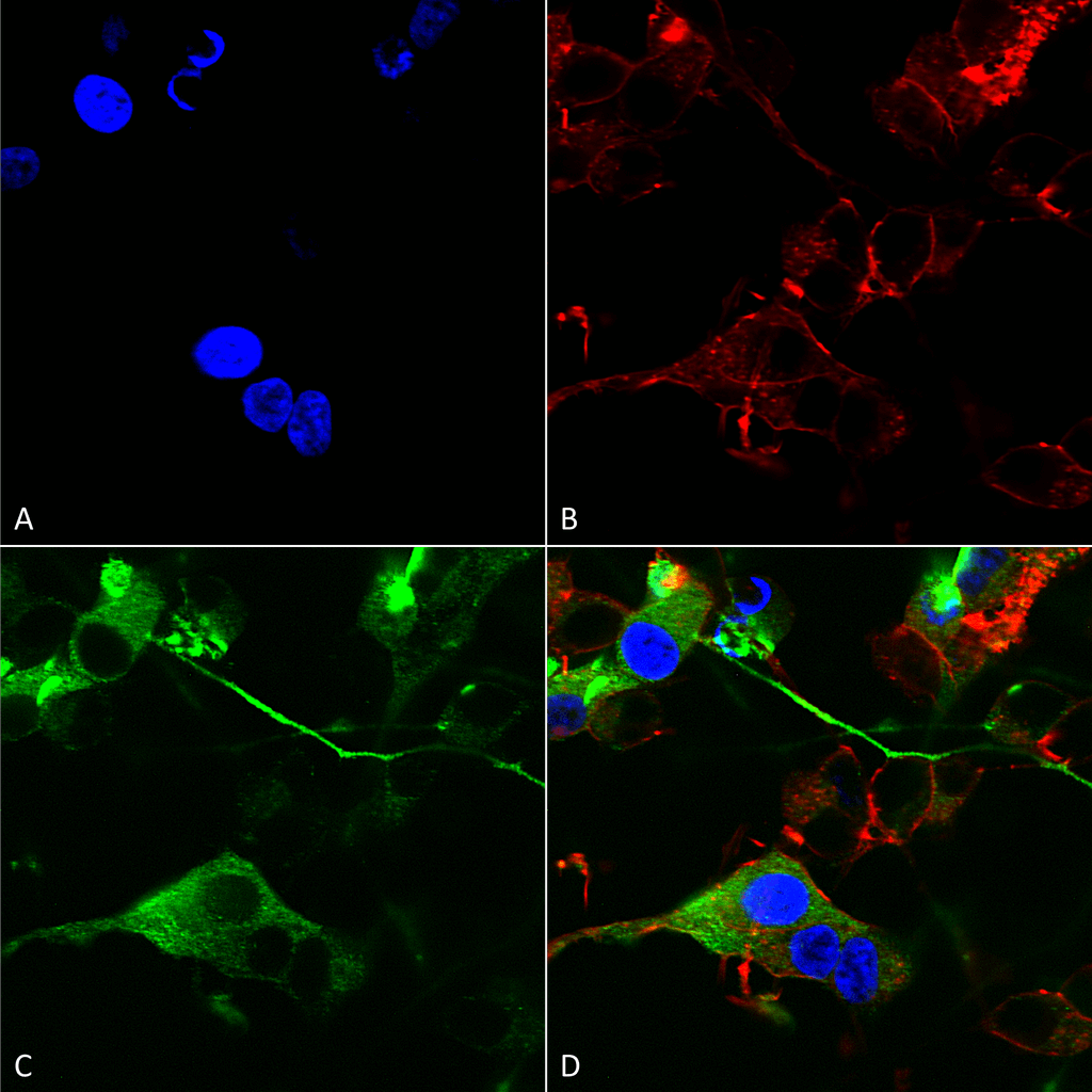
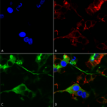
Immunocytochemistry/Immunofluorescence analysis using Mouse Anti-GABA-A Receptor Alpha 2 Monoclonal Antibody, Clone N399/19 (SMC-486). Tissue: Neuroblastoma cells (SH-SY5Y). Species: Human. Fixation: 4% PFA for 15 min. Primary Antibody: Mouse Anti-GABA-A Receptor Alpha 2 Monoclonal Antibody (SMC-486) at 1:200 for overnight at 4°C with slow rocking. Secondary Antibody: AlexaFluor 488 at 1:1000 for 1 hour at RT. Counterstain: Phalloidin-iFluor 647 (red) F-Actin stain; Hoechst (blue) nuclear stain at 1:800, 1.6mM for 20 min at RT. (A) Hoechst (blue) nuclear stain. (B) Phalloidin-iFluor 647 (red) F-Actin stain. (C) GABA-A Receptor Alpha 2 Antibody (D) Composite.
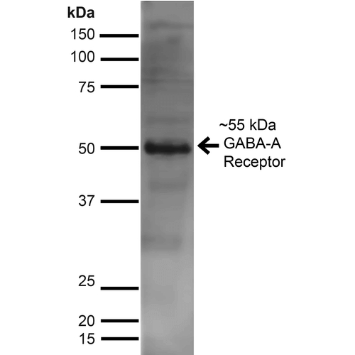
Western Blot analysis of Rat Brain showing detection of ~55 kDa GABA A Receptor Alpha 2 protein using Mouse Anti-GABA A Receptor Alpha 2 Monoclonal Antibody, Clone N399/19 (SMC-486). Lane 1: MW Ladder. Lane 2: Rat Brain. Load: 10 µg. Block: 5% Skim Milk for 1 hour at RT. Primary Antibody: Mouse Anti-GABA A Receptor Alpha 2 Monoclonal Antibody (SMC-486) at 1:1000 for 1 hour at RT. Secondary Antibody: Goat Anti-Mouse IgG: HRP at 1:100 for 1 hour at RT. Color Development: ECL solution for 6 min at RT. Predicted/Observed Size: ~55 kDa.
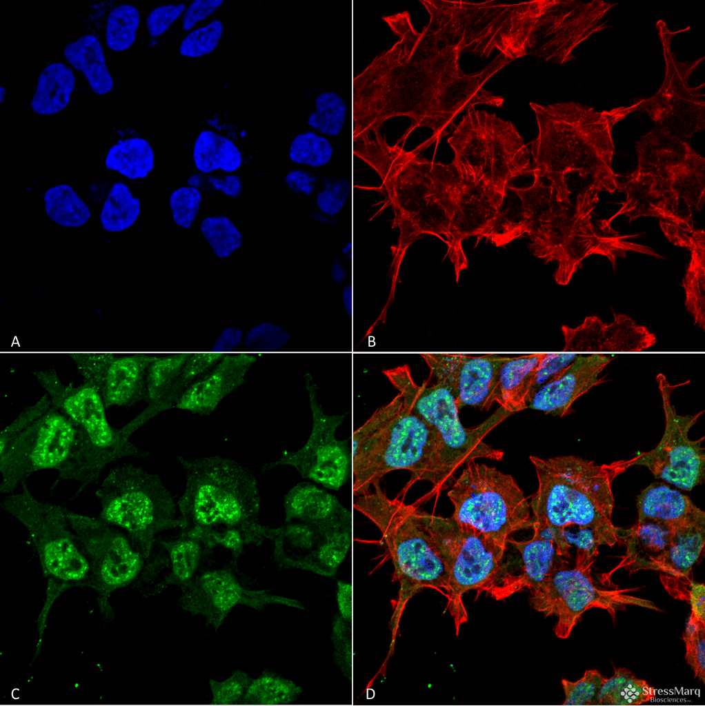
Immunocytochemistry/Immunofluorescence analysis using Mouse Anti-GABA-A Receptor Alpha2 Monoclonal Antibody, Clone N399/19 (SMC-486). Tissue: Neuroblastoma cell line (SK-N-BE). Species: Human. Fixation: 4% Formaldehyde for 15 min at RT. Primary Antibody: Mouse Anti-GABA-A Receptor Alpha2 Monoclonal Antibody (SMC-486) at 1:100 for 60 min at RT. Secondary Antibody: Goat Anti-Mouse ATTO 488 at 1:100 for 60 min at RT. Counterstain: Phalloidin Texas Red F-Actin stain; DAPI (blue) nuclear stain at 1:1000; 1:5000 for 60 min RT, 5 min RT. Localization: Cytoplasm, Nucleus. Magnification: 60X. (A) DAPI (blue) nuclear stain. (B) Phalloidin Texas Red F-Actin stain. (C) GABA-A Receptor Alpha2 Antibody. (D) Composite.
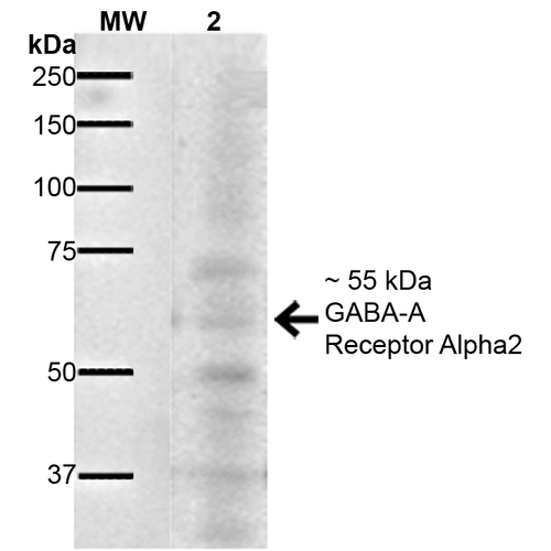
Western Blot analysis of Mouse Brain showing detection of ~55 kDa GABA A Receptor Alpha 2 protein using Mouse Anti-GABA A Receptor Alpha 2 Monoclonal Antibody, Clone N399/19 (SMC-486). Lane 1: MW Ladder. Lane 2: Mouse Brain. Load: 20 µg. Primary Antibody: Mouse Anti-GABA A Receptor Alpha 2 Monoclonal Antibody (SMC-486) at 1:1000 for 16 hours at 4°C. Secondary Antibody: Goat Anti-Mouse IgG: HRP at 1:200 for 1 hour at RT. Predicted/Observed Size: ~55 kDa. Other Band(s): ~ 37 kDa, ~50 kDa, ~70 kDa.



Reviews
There are no reviews yet.