Properties
| Storage Buffer | PBS pH7.4, 50% glycerol, 0.09% sodium azide *Storage buffer may change when conjugated |
| Storage Temperature | -20ºC, Conjugated antibodies should be stored according to the product label |
| Shipping Temperature | Blue Ice or 4ºC |
| Purification | Protein G Purified |
| Clonality | Monoclonal |
| Clone Number | N81/2 (Formerly sold as S81-2) |
| Isotype | IgG1 |
| Specificity | Detects ~105kDa. No cross-reactivity against GABA(B)R1. |
| Cite This Product | GABAB Receptor 2 Antibody (StressMarq Biosciences | Victoria, BC CANADA, Catalog# SMC-402, RRID: AB_11232611) |
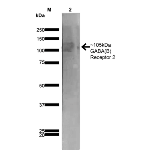
| Certificate of Analysis | 1 µg/ml of SMC-402 was sufficient for detection of GABA(B)R2 in 20 µg of rat brain membrane lysate and assayed by colorimetric immunoblot analysis using goat anti-mouse IgG:HRP as the secondary antibody. |
Biological Description
| Alternative Names | GABA B receptor 2, GABA-B receptor 2, GABA-B-R2, GABA-BR2, GABAB R2, GABABR2, GABBR 2, GABBR2, GABR2_HUMAN, Gamma aminobutyric acid (GABA) B receptor 2, Gamma aminobutyric acid B receptor 2, Gamma aminobutyric acid GABA B receptor 2, Gamma aminobutyric acid type B receptor subunit 2, Gb 2, Gb2, GPR 51, GPR51, G protein coupled receptor 51, G-protein coupled receptor 51, GPRC 3B, GPRC3B, HG 20, HG20, HRIHFB2099, Metabotropic GABA B receptor subtype 2, BcDNA:GH07312, CG6706, CT20836, D GABA[[B]]R2, D Gaba2, FLJ36928, GH07312, OTTHUMP00000021776, OTTHUMP00000063797, R2 SUBUNIT |
| Research Areas | GABA Receptors, GABAB Receptors, Neuroscience, Neurotransmitter Receptors |
| Cellular Localization | Cell Junction, Cell membrane, Postsynaptic cell membrane, Synapse |
| Accession Number | NP_113990.1 |
| Gene ID | 83633 |
| Swiss Prot | O88871 |
| Scientific Background |
GABA (γ-aminobutyric acid) is the primary inhibitory neurotransmitter in the central nervous system, acting through three receptor types: GABA-A, GABA-B, and GABA-C. While GABA-A and GABA-C receptors are ionotropic and mediate rapid synaptic inhibition, GABA-B receptors are metabotropic, G protein-coupled receptors that regulate slower, prolonged inhibitory signaling. Functional GABA-B receptors are obligate heterodimers composed of GABA-B Receptor 1 (GABBR1), which binds GABA, and GABA-B Receptor 2 (GABBR2), which is essential for G protein coupling and intracellular signaling. GABBR2 plays a pivotal role in activating downstream pathways that inhibit adenylate cyclase, suppress voltage-gated calcium channels, and activate inwardly rectifying potassium channels. These mechanisms contribute to both presynaptic inhibition—by reducing neurotransmitter release—and postsynaptic inhibition—by hyperpolarizing neurons. Beyond its role in synaptic inhibition, GABBR2 is involved in regulating hippocampal long-term potentiation, sleep architecture, and motor control. Dysregulation of GABBR2 has been increasingly implicated in neurodegenerative diseases such as Alzheimer’s, Parkinson’s, and Huntington’s disease. Altered GABBR2 expression or function may disrupt inhibitory tone, contribute to excitotoxicity, and impair synaptic plasticity—key features of neurodegeneration. As a result, GABBR2 is emerging as a promising therapeutic target and biomarker in neuroscience, offering new opportunities for modulating GABAergic signaling to preserve cognitive and neural function in aging and disease. |
| References |
1. Jones K.A,. et al. (2000) Neuropsychopharmacology 23: S41-9. 2. Duthey B., et al. (2002) J Biol Chem. 277: 3236-41. 3. Kaupmann K., et al. (1997) Nature 386: 239-46. |
Product Images

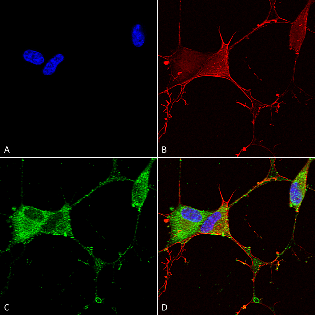
Immunocytochemistry/Immunofluorescence analysis using Mouse Anti-GABA-B Receptor 2 Monoclonal Antibody, Clone N81/2 (SMC-402). Tissue: Neuroblastoma cells (SH-SY5Y). Species: Human. Fixation: 4% PFA for 15 min. Primary Antibody: Mouse Anti-GABA-B Receptor 2 Monoclonal Antibody (SMC-402) at 1:100 for overnight at 4°C with slow rocking. Secondary Antibody: AlexaFluor 488 at 1:1000 for 1 hour at RT. Counterstain: Phalloidin-iFluor 647 (red) F-Actin stain; Hoechst (blue) nuclear stain at 1:800, 1.6mM for 20 min at RT. (A) Hoechst (blue) nuclear stain. (B) Phalloidin-iFluor 647 (red) F-Actin stain. (C) GABA-B Receptor 2 Antibody (D) Composite.

Western Blot analysis of Rat Brain Membrane showing detection of ~105 kDa GABA B Receptor 2 protein using Mouse Anti-GABA B Receptor 2 Monoclonal Antibody, Clone N81/2 (SMC-402). Lane 1: MW Ladder. Lane 2: Rat Brain Membrane (10 µg). . Load: 10 µg. Block: 5% milk. Primary Antibody: Mouse Anti-GABA B Receptor 2 Monoclonal Antibody (SMC-402) at 1:1000 for 1 hour at RT. Secondary Antibody: Goat Anti-Mouse IgG: HRP at 1:200 for 1 hour at RT. Color Development: TMB solution for 10 min at RT. Predicted/Observed Size: ~105 kDa.

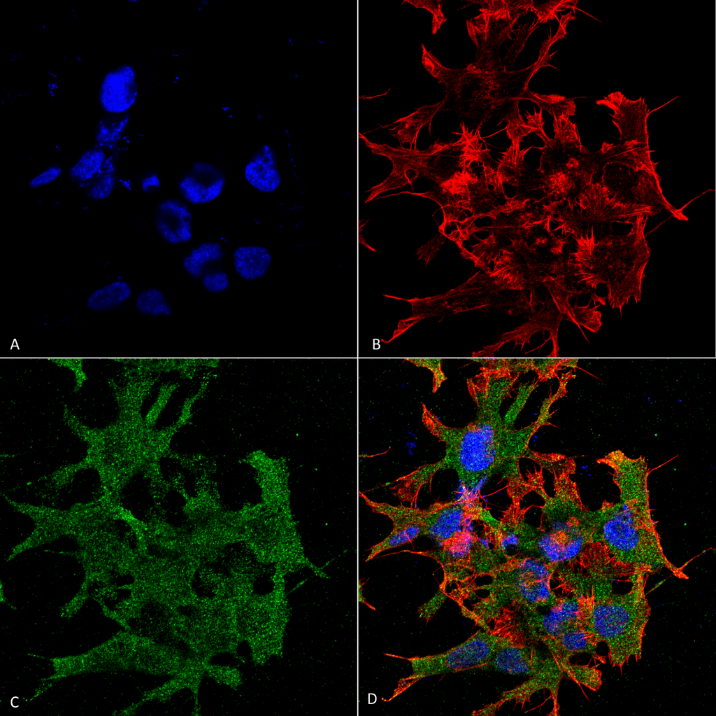
Immunocytochemistry/Immunofluorescence analysis using Mouse Anti-GABA-B Receptor 2 Monoclonal Antibody, Clone N81/2 (SMC-402). Tissue: Neuroblastoma cell line (SK-N-BE). Species: Human. Fixation: 4% Formaldehyde for 15 min at RT. Primary Antibody: Mouse Anti-GABA-B Receptor 2 Monoclonal Antibody (SMC-402) at 1:100 for 60 min at RT. Secondary Antibody: Goat Anti-Mouse ATTO 488 at 1:100 for 60 min at RT. Counterstain: Phalloidin Texas Red F-Actin stain; DAPI (blue) nuclear stain at 1:1000, 1:5000 for 60min RT, 5min RT. Localization: Cell Membrane. Magnification: 60X. (A) DAPI (blue) nuclear stain. (B) Phalloidin Texas Red F-Actin stain. (C) GABA-B Receptor 2 Antibody. (D) Composite.


Reviews
There are no reviews yet.