Discovery through Partnership | Excellence through Quality


Properties
| Storage Buffer | PBS pH 7.4, 50% glycerol, 0.09% Sodium azide *Storage buffer may change when conjugated |
| Storage Temperature | -20ºC, Conjugated antibodies should be stored according to the product label |
| Shipping Temperature | Blue Ice or 4ºC |
| Purification | Protein G Purified |
| Clonality | Recombinant Monoclonal |
| Clone Number | 1H11 |
| Isotype | IgG1 |
| Specificity | Conformation-specific antibody for membrane-bound HSP70. Binds to cell surface HSP70 without permeabilization of cell membrane in tumor cell lines. Does not bind to intracellular HSP70 at low concentrations. Cross-reacts with HSC70. Detects a ~70 kDa band in Western Blot. |
| Cite This Product | HSP70 Antibody (StressMarq Biosciences | Victoria, BC CANADA, Catalog# SMC-249, RRID: AB_2699628) |
| Certificate of Analysis | A 1:250 dilution of SMC-249 was sufficient for detection of cell surface HSP70 in HCT116 cells using Fluorescence-activated cell sorting, applied in flow cytometry, with FITC as the fluorescent probe. |
Biological Description
| Alternative Names | HSPA1A, HSPA1B, HSPA1, HSP70, HSP70-1, HSP70.1, HSP70-2, HSP72, HSP73, HSX70, Heat shock 70 kDa protein 1A, Heat shock 70 kDa protein 1B |
| Research Areas | Cancer, Cell Signaling, Chaperone Proteins, Heat Shock, Protein Trafficking, Tumor Biomarkers |
| Cellular Localization | Cell membrane |
| Accession Number | NP_005336.3 |
| Gene ID | 3303 |
| Swiss Prot | P0DMV8/P0DMV9 |
| Scientific Background |
HSP70 proteins are a highly conserved family of 70-kDa molecular chaperones encoded by a multigene family in most eukaryotes and prokaryotes. Found in nearly all cellular compartments—including the cytosol, nucleus, mitochondria, endoplasmic reticulum, and chloroplasts—HSP70s are constitutively expressed and strongly upregulated in response to cellular stress. These chaperones play a vital role in protein homeostasis by binding to nascent polypeptides and partially folded or misfolded proteins, preventing aggregation and facilitating proper folding. HSP70s exhibit high-affinity ATP binding and weak ATPase activity, which is stimulated upon interaction with unfolded substrates. ATP hydrolysis triggers conformational changes that regulate substrate binding and release, enabling dynamic cycles of protein folding and refolding. Structurally, the N-terminal domain of HSP70 is responsible for ATP binding, while the C-terminal domain mediates substrate interaction. This modular design allows HSP70s to coordinate with co-chaperones such as HSP40, HIP, HOP, and BAG-1, integrating folding with degradation and transport pathways. In neurodegenerative disease research, HSP70 is of particular interest due to its ability to counteract protein misfolding and aggregation—hallmarks of disorders like Alzheimer’s, Parkinson’s, and Huntington’s disease. By stabilizing toxic intermediates and promoting their clearance, HSP70 enhances neuronal survival and resilience under proteotoxic stress. |
| References |
1. Gribaldo, S. et al. (1999) J. Bacteriol. 181, 434-443. 2. Sharma, D. & Masison, D.C. (2009) Protein Pept. Lett. 16, 571-581. 3. Sharma, D. et al. (2009) PLoS. ONE.4, e6644. 4. Kampinga, H.H. & Craig, E.A. (2010) Nat. Rev. Mol. Cell Biol. 11, 579-592. 5. Nickel, W. & Seedorf, M. (2008) Annu. Rev. Cell Dev. Biol. 24, 287-308. 6. Multhoff, G. & Hightower, L.E. (1996) Cell Stress. Chaperones. 1, 167-176. 7. De Maio, A. (2011) Cell Stress. Chaperones. 16, 235-249. 8. Horvath, I. & Vigh, L. (2010) Nature. 463, 436-438. 9. Horvath, I., Multhoff, G., Sonnleitner, A., & Vigh, L. (2008) Biochim. Biophys. Acta 1778, 1653-1664. |
Product Images
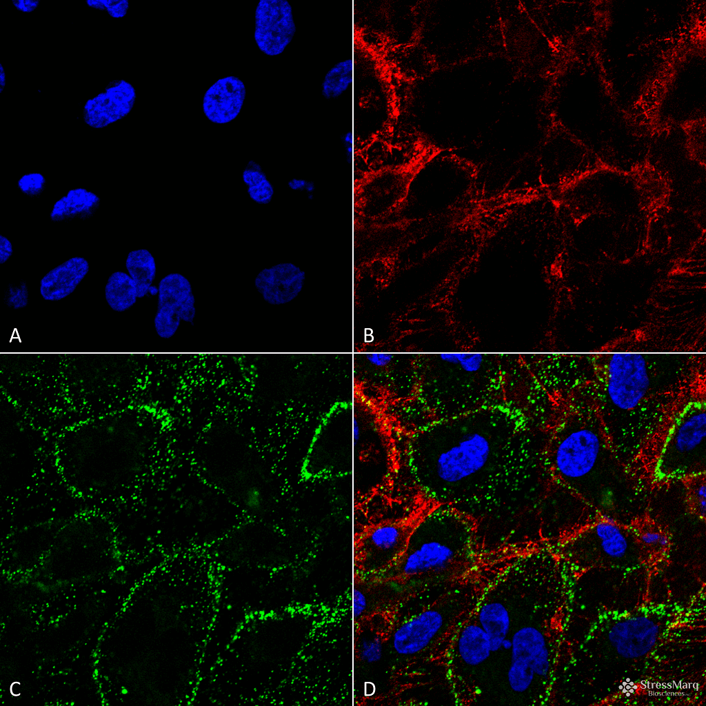
Immunocytochemistry/Immunofluorescence analysis using Mouse Anti-HSP70 Monoclonal Antibody, Clone 1H11 (SMC-249). Tissue: HCT116 cells. Species: Human. Fixation: 4% Formaldehyde. Primary Antibody: Mouse Anti-HSP70 Monoclonal Antibody (SMC-249) at 1:100. Counterstain: Wheat germ agglutinin Texas red membrane marker; DAPI (blue) nuclear stain. Localization: Cell surface, cell membrane. (A) DAPI nuclear stain. (B) Wheat germ agglutinin Texas red. (C) HSP70 Antibody. (D) Composite. Courtesy of: Lawrence Hightower, Charles Giardina, and Didem Ozcan from University of Connecticut.
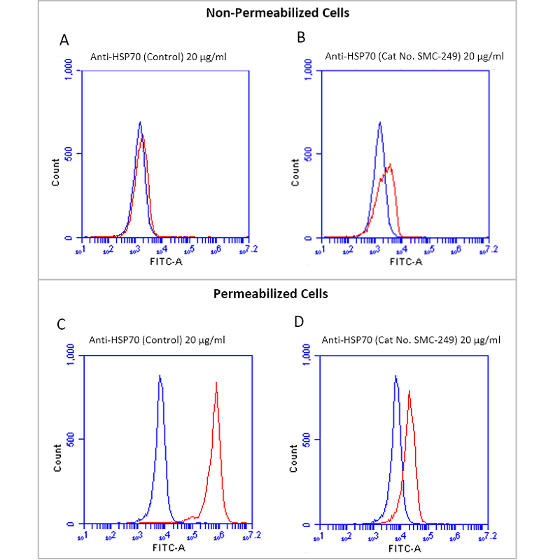
Fluorescence-activated cell sorting analysis using Mouse Anti-HSP70 Monoclonal Antibody, Clone 1H11 (SMC-249). Tissue: Jurkat E6.1 cells. Species: Human. Fixation: No fixation. Primary Antibody: Mouse Anti-HSP70 Monoclonal Antibody (SMC-249) at 20 µg/ml for 40 min at 4°C. Counterstain: Propidium Iodide nuclear stain at 2.5 µg/ml for 5 min at RT. Isotype Control: Anti-mouse FITC at 1:32 for 15 min at RT (blue line). Courtesy of: Dr. Elyse Ireland, Institute of Medicine, University of Chester.
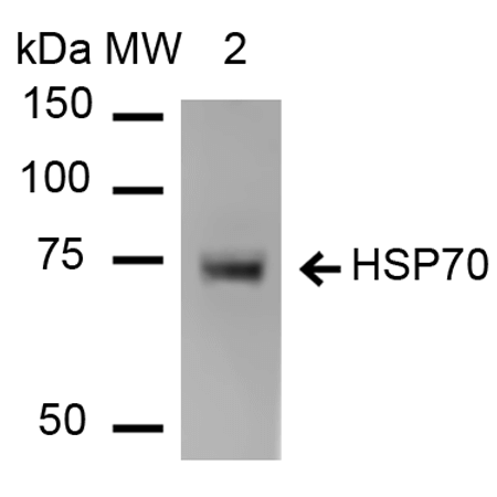
Western Blot analysis of Human Heat Shocked cervical cancer cell line (HeLa) lysate showing detection of HSP70 protein using Mouse Anti-HSP70 Monoclonal Antibody, Clone 1H11 (SMC-249). Lane 1: Molecular Weight ladder (MW). Lane 2: HeLa cell lysates. Load: 20 µg. Primary Antibody: Mouse Anti-HSP70 Monoclonal Antibody (SMC-249) at 1:1000.
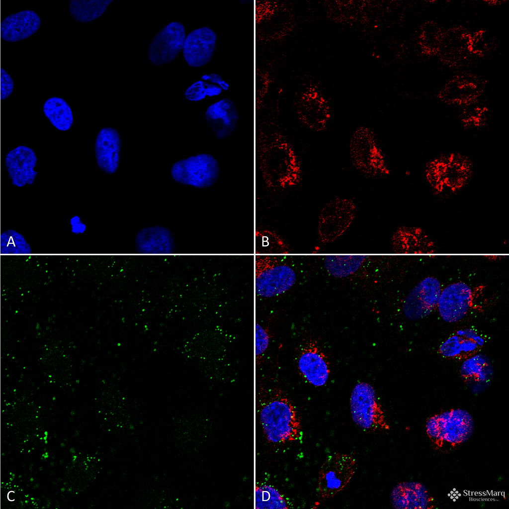
Immunocytochemistry/Immunofluorescence analysis using Mouse Anti-HSP70 Monoclonal Antibody, Clone 1H11 (SMC-249). Tissue: HCT116 cells. Species: Human. Fixation: 4% Formaldehyde. Primary Antibody: Mouse Anti-HSP70 Monoclonal Antibody (SMC-249) at 1:100. Counterstain: Wheat germ agglutinin Texas red membrane marker; DAPI (blue) nuclear stain. Localization: Cell surface, faint intracellular. (A) DAPI nuclear stain. (B) Wheat germ agglutinin Texas red. (C) HSP70 Antibody. (D) Composite. Courtesy of: Lawrence Hightower, Charles Giardina, and Didem Ozcan from University of Connecticut.
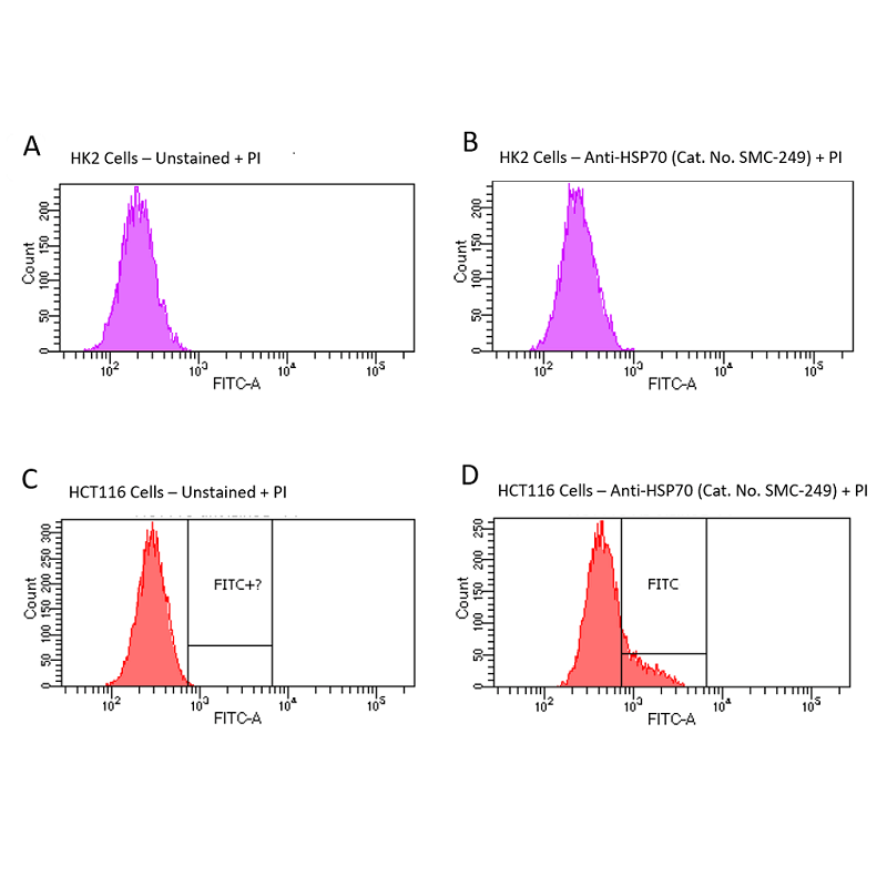
Fluorescence-activated cell sorting analysis using Mouse Anti-HSP70 Monoclonal Antibody, Clone 1H11 (SMC-249). Tissue: HCT116 and HK2 cells. Species: Human. Primary Antibody: Mouse Anti-HSP70 Monoclonal Antibody (SMC-249) at 1:100 for 90 min at 4°C. Counterstain: Propidium Iodide nuclear stain. (A) HK2 cells unstained. (B) HK2 cells and HSP70 Antibody. (C) HCT116 cells unstained. (D) HCT116 cells and HSP70 Antibody. Shows that HSP70 Antibody binds to the cell surface of tumor cells but not non-tumor cells. Courtesy of: Lawrence Hightower, Charles Giardina, and Didem Ozcan from University of Connecticut.
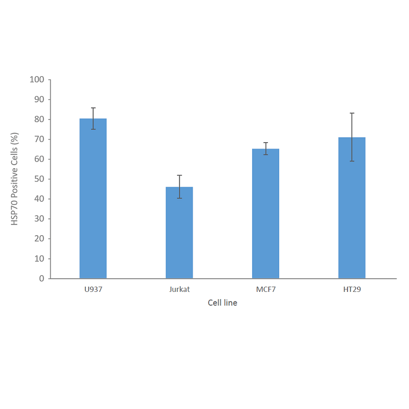
Fluorescence-activated cell sorting analysis using Mouse Anti-HSP70 Monoclonal Antibody, Clone 1H11 (SMC-249). Tissue: Jurkat E6.1, U937, MCF7 and HT29 cells. Species: Human. Fixation: No fixation. Primary Antibody: Mouse Anti-HSP70 Monoclonal Antibody (SMC-249) at 20 µg/ml for 40 min at 4°C. Counterstain: Propidium Iodide nuclear stain at 2.5 µg/ml for 5 min at 4°C. Shows that binding of HSP70 Antibody is not cell-line specific. Courtesy of: Dr. Elyse Ireland, Institute of Medicine, University of Chester.
 Powered by Bioz
Samantha Chin :
High-quality antibody with ideal clone for tumor cell studies – great signal on the cytometer. See full review: https://www.biocompare.com/Product-Reviews/598227-High-quality-antibody-for-flow-cytometry/#disqus_thread
StressMarq Biosciences :
Based on validation through cited publications.