Discovery through Partnership | Excellence through Quality


Properties
| Storage Buffer | PBS pH7.4, 50% glycerol, 0.09% sodium azide *Storage buffer may change when conjugated |
| Storage Temperature | -20ºC, Conjugated antibodies should be stored according to the product label |
| Shipping Temperature | Blue Ice or 4ºC |
| Purification | Protein G Purified |
| Clonality | Monoclonal |
| Clone Number | N37A/10 (Formerly sold as S37A-10) |
| Isotype | IgG1 |
| Specificity | Detects ~75kDa. |
| Cite This Product | KCNQ1 Antibody (StressMarq Biosciences | Victoria, BC CANADA, Catalog# SMC-307, RRID: AB_2249571) |
| Certificate of Analysis | 1 µg/ml of SMC-307 was sufficient for detection of KCNQ1 in 10 µg of COS-1 cell lysate transiently expressing KCNQ1 by colorimetric immunoblot analysis using Goat anti-mouse IgG:HRP as the secondary antibody. |
Biological Description
| Alternative Names | KCNQ1, Kv7.1, KVLQT1, Potassium voltage-gated channel subfamily KQT member 1, Voltage-gated potassium channel subunit Kv7.1, KQT-like 1, IKs producing slow voltage-gated potassium channel subunit alpha, Slow delayed rectifier channel subunit, ATFB1, ATFB3, FLJ26167, JLNS1, KCNA8, KCNA9, KCNQ1_HUMAN, Kidney and cardiac voltage dependent K+ channel, Kv1.9, Long (electrocardiographic) QT syndrome, LQT, LQT1, RWS, SQT2, Ward-Romano syndrome 1 |
| Research Areas | Cardiovascular System, Heart, Ion Channels, Neuroscience, Potassium Channels, Voltage-Gated Potassium Channels |
| Cellular Localization | Cell membrane, Cytoplasmic vesicle membrane |
| Accession Number | NP_000209.2 |
| Gene ID | 3784 |
| Swiss Prot | P51787 |
| Scientific Background |
KCNQ1 encodes the Kv7.1 potassium channel, a voltage-gated protein best known for its role in cardiac electrophysiology. In cardiac myocytes, Kv7.1 mediates the slow delayed rectifier potassium current (IKs), which is essential for repolarization and termination of the cardiac action potential. However, its expression in non-cardiac tissues, including inner ear neurons and select brain regions, suggests broader physiological relevance. Recent research has begun to uncover Kv7.1’s potential role in the nervous system, particularly in regulating neuronal excitability and maintaining ionic homeostasis. Potassium channels like Kv7.1 are critical for shaping action potentials, modulating synaptic transmission, and protecting neurons from hyperexcitability and excitotoxicity—key features implicated in neurodegenerative disease progression. Although less studied than other Kv7 family members in the brain, KCNQ1’s presence in neural tissue raises important questions about its contribution to auditory processing, neurodevelopment, and neurodegeneration. Dysregulation of potassium channel activity has been linked to disorders such as epilepsy, Alzheimer’s disease, and Parkinson’s disease, where altered ion channel function disrupts neural signaling and accelerates neuronal loss. As neuroscience research expands its focus on ion channelopathies, KCNQ1 is emerging as a candidate for further investigation. Understanding its role in neural circuits may reveal novel therapeutic strategies for restoring electrical balance and preventing neurodegenerative decline. |
| References |
1. Lang F., Vallon V., Knipper M., Wagenmann P. (2007) Am J Physiol. 293(4): C1187-1208. 2. Kurokawa J., et al. (2009) Channels (Austin). 3(1): 16-24. 3. Silva J., and Rudy Y. (2005) Circulation. 112(10): 1384-1391. |
Product Images
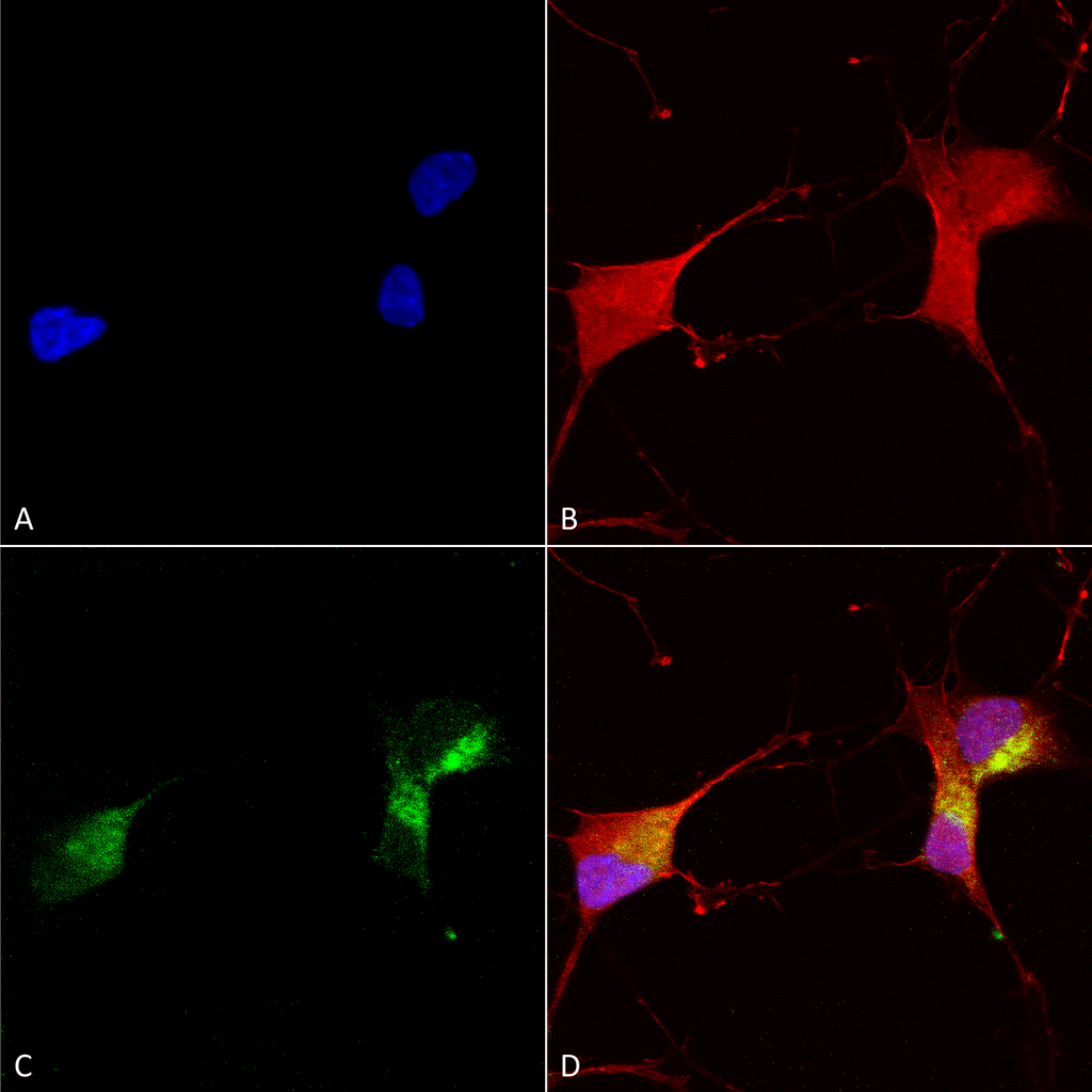
Immunocytochemistry/Immunofluorescence analysis using Mouse Anti-KCNQ1 Monoclonal Antibody, Clone N37A/10 (SMC-307). Tissue: Neuroblastoma cells (SH-SY5Y). Species: Human. Fixation: 4% PFA for 15 min. Primary Antibody: Mouse Anti-KCNQ1 Monoclonal Antibody (SMC-307) at 1:100 for overnight at 4°C with slow rocking. Secondary Antibody: AlexaFluor 488 at 1:1000 for 1 hour at RT. Counterstain: Phalloidin-iFluor 647 (red) F-Actin stain; Hoechst (blue) nuclear stain at 1:800, 1.6mM for 20 min at RT. (A) Hoechst (blue) nuclear stain. (B) Phalloidin-iFluor 647 (red) F-Actin stain. (C) KCNQ1 Antibody (D) Composite.
Immunohistochemistry analysis using Mouse Anti-KCNQ1 Monoclonal Antibody, Clone N37A/10 (SMC-307). Tissue: hippocampus. Species: Human. Fixation: Bouin’s Fixative and paraffin-embedded. Primary Antibody: Mouse Anti-KCNQ1 Monoclonal Antibody (SMC-307) at 1:1000 for 1 hour at RT. Secondary Antibody: FITC Goat Anti-Mouse (green) at 1:50 for 1 hour at RT.
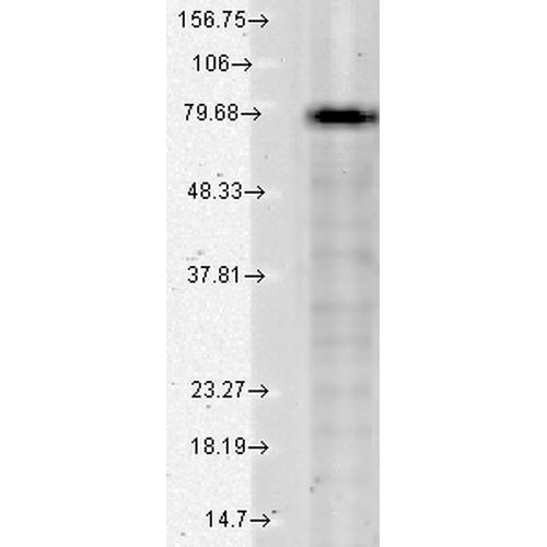
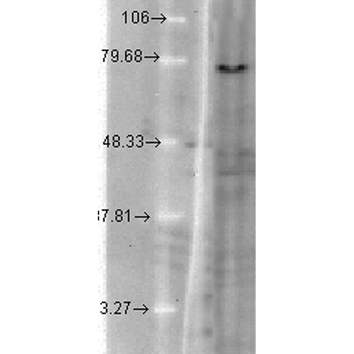
Western Blot analysis of Human Cell lysates showing detection of KCNQ1 protein using Mouse Anti-KCNQ1 Monoclonal Antibody, Clone N37A/10 (SMC-307). Load: 15 µg. Block: 1.5% BSA for 30 minutes at RT. Primary Antibody: Mouse Anti-KCNQ1 Monoclonal Antibody (SMC-307) at 1:1000 for 2 hours at RT. Secondary Antibody: Sheep Anti-Mouse IgG: HRP for 1 hour at RT.
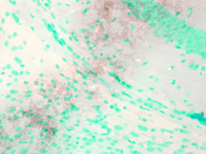
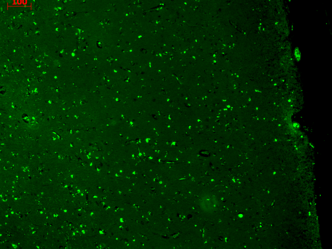
Immunohistochemistry analysis using Mouse Anti-KCNQ1 Monoclonal Antibody, Clone N37A/10 (SMC-307). Tissue: Brain Slice. Species: Mouse. Fixation: 10% Formalin Solution for 12-24 hours at RT. Primary Antibody: Mouse Anti-KCNQ1 Monoclonal Antibody (SMC-307) at 1:1000 for 1 hour at RT. Secondary Antibody: HRP/DAB Detection System: Biotinylated Goat Anti-Mouse, Streptavidin Peroxidase, DAB Chromogen (brown) for 30 minutes at RT. Counterstain: Mayer Hematoxylin (purple/blue) nuclear stain at 250-500 µl for 5 minutes at RT.
Western Blot analysis of Hamster T-CHO cell lysate showing detection of KCNQ1 protein using Mouse Anti-KCNQ1 Monoclonal Antibody, Clone N37A/10 (SMC-307). Load: 15 µg. Block: 1.5% BSA for 30 minutes at RT. Primary Antibody: Mouse Anti-KCNQ1 Monoclonal Antibody (SMC-307) at 1:1000 for 2 hours at RT. Secondary Antibody: Sheep Anti-Mouse IgG: HRP for 1 hour at RT.
 Powered by Bioz
StressMarq Biosciences :
Based on validation through cited publications.