Discovery through Partnership | Excellence through Quality


Properties
| Storage Buffer | PBS pH 7.4, 50% glycerol, 0.09% Sodium azide *Storage buffer may change when conjugated |
| Storage Temperature | -20ºC, Conjugated antibodies should be stored according to the product label |
| Shipping Temperature | Blue Ice or 4ºC |
| Purification | Protein G Purified |
| Clonality | Monoclonal |
| Clone Number | 11E3 |
| Isotype | IgG1 |
| Specificity | Specific for Malondialdehyde conjugated proteins. Does not detect free Malondialdehyde. Does not cross-react with Acrolein, Crotonaldehyde, Hexanoyl Lysine, 4-Hydroxy-2-hexenal, 4-Hydroxy nonenal, or Methylglyoxal modified proteins. |
| Cite This Product | Malondialdehyde Antibody (StressMarq Biosciences | Victoria, BC CANADA, Catalog# SMC-515, RRID: AB_2702909) |
| Certificate of Analysis | A 1:1000 dilution of SMC-515 was sufficient for detection of Malondialdehyde in 2 µg of Malondialdehyde conjugated to BSA by ECL immunoblot analysis using Goat Anti-Mouse IgG:HRP as the secondary Antibody. |
Biological Description
| Alternative Names | Malondialdehyde, MDA, Malondialdehyde (MDA), Malonic aldehyde, Propanedial, 1,3-Propanedial, Malonaldehyde |
| Research Areas | Alzheimer's Disease, Cancer, Lipid peroxidation, Neurodegeneration, Neuroscience, Oxidative Stress |
| Scientific Background |
Malondialdehyde (MDA) is a highly reactive, low-molecular-weight aldehyde and one of the most widely used biomarkers of oxidative stress due to its chemical stability and ease of detection. Formed as a byproduct of lipid peroxidation, MDA reflects oxidative damage to polyunsaturated fatty acids in cellular membranes—a process central to the pathology of neurodegenerative diseases. MDA exhibits a strong affinity for amino groups, forming adducts with free amino acids, proteins, and nucleic acids. These adducts can disrupt protein function and induce DNA cross-linking, contributing to mutagenesis and cellular dysfunction. Elevated MDA levels have been consistently observed in Alzheimer’s disease and other neurodegenerative conditions, as well as in systemic diseases such as diabetes and cancer. In neuroscience research, MDA serves not only as a diagnostic and prognostic marker but also as a mechanistic link between oxidative stress and neuronal injury. Its quantification—commonly performed using the thiobarbituric acid reactive substances (TBARS) assay—provides valuable insights into the redox state of the brain and the efficacy of antioxidant therapies. Given its dual role as both a biomarker and a mediator of oxidative damage, MDA is a critical target in neurodegenerative disease research. Understanding and mitigating MDA-associated toxicity may offer new avenues for therapeutic intervention in oxidative stress-driven neurological disorders. |
Product Images
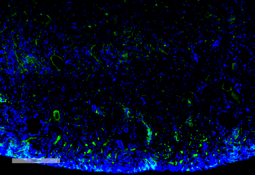
Immunohistochemistry analysis using Mouse Anti-Malondialdehyde Monoclonal Antibody, Clone 11E3 (SMC-515). Tissue: Kidney. Species: Mouse. Primary Antibody: Mouse Anti-Malondialdehyde Monoclonal Antibody (SMC-515) at 1:100 for Overnight at 4C, then 30 min at 37C. Secondary Antibody: Goat Anti-Mouse IgG (H+L): FITC for 45 min at 37C. Counterstain: DAPI for 3 min at RT. Magnification: 10X.
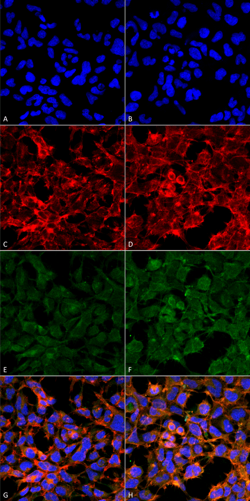
Immunocytochemistry/Immunofluorescence analysis using Mouse Anti-Malondialdehyde Monoclonal Antibody, Clone 11E3 (SMC-515). Tissue: Embryonic kidney epithelial cell line (HEK293). Species: Human. Fixation: 5% Formaldehyde for 5 min. Primary Antibody: Mouse Anti-Malondialdehyde Monoclonal Antibody (SMC-515) at 1:50 for 30-60 min at RT. Secondary Antibody: Goat Anti-Mouse Alexa Fluor 488 at 1:1500 for 30-60 min at RT. Counterstain: Phalloidin Alexa Fluor 633 F-Actin stain; DAPI (blue) nuclear stain at 1:250, 1:50000 for 30-60 min at RT. Magnification: 20X (2X Zoom). (A,C,E,G) – Untreated. (B,D,F,H) – Cells cultured overnight with 50 µM H2O2. (A,B) DAPI (blue) nuclear stain. (C,D) Phalloidin Alexa Fluor 633 F-Actin stain. (E,F) Malondialdehyde Antibody. (G,H) Composite. Courtesy of: Dr. Robert Burke, University of Victoria.
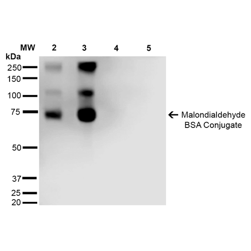
Western Blot analysis of Malondialdehyde-BSA Conjugate showing detection of 67 kDa Malondialdehyde protein using Mouse Anti-Malondialdehyde Monoclonal Antibody, Clone 11E3 (SMC-515). Lane 1: Molecular Weight Ladder (MW). Lane 2: Malondialdehyde-BSA (0.5 µg). Lane 3: Malondialdehyde-BSA (2.0 µg). Lane 4: BSA (0.5 µg). Lane 5: BSA (2.0 µg) . Block: 5% Skim Milk in TBST. Primary Antibody: Mouse Anti-Malondialdehyde Monoclonal Antibody (SMC-515) at 1:1000 for 2 hours at RT. Secondary Antibody: Goat Anti-Mouse IgG: HRP at 1:2000 for 60 min at RT. Color Development: ECL solution for 5 min in RT. Predicted/Observed Size: 67 kDa.
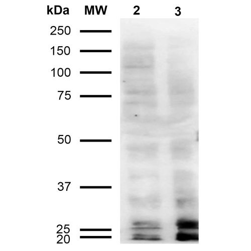
Western Blot analysis of Human Cervical cancer cell line (HeLa) lysate showing detection of Malondialdehyde protein using Mouse Anti-Malondialdehyde Monoclonal Antibody, Clone 11E3 (SMC-515). Lane 1: Molecular Weight Ladder (MW). Lane 2: HeLa cell lysate. Lane 3: H2O2 treated HeLa cell lysate. Load: 12 µg. Block: 5% Skim Milk in TBST. Primary Antibody: Mouse Anti-Malondialdehyde Monoclonal Antibody (SMC-515) at 1:1000 for 2 hours at RT. Secondary Antibody: Goat Anti-Mouse IgG: HRP at 1:2000 for 60 min at RT. Color Development: ECL solution for 5 min in RT.
 Powered by Bioz
Reviews
There are no reviews yet.