Discovery through Partnership | Excellence through Quality


Properties
| Storage Buffer | PBS pH 7.4, 50% glycerol, 0.09% Sodium azide *Storage buffer may change when conjugated |
| Storage Temperature | -20ºC, Conjugated antibodies should be stored according to the product label |
| Shipping Temperature | Blue Ice or 4ºC |
| Purification | Protein G Purified |
| Clonality | Monoclonal |
| Clone Number | 1D5 |
| Isotype | IgG1 |
| Specificity | Detects Multiple Bands. Antibody detects monomer under denaturing conditions but preferentially detects fibril under native conditions (dot blot). |
| Cite This Product | Tau Antibody (StressMarq Biosciences | Victoria, BC CANADA, Catalog# SMC-607, RRID: AB_2820306) |
| Certificate of Analysis | A 1:1000 dilution of SMC-607 was sufficient for detection of Tau 2N4R P301S Fibril in 20 ug of Mouse Brain by ECL immunoblot analysis using goat anti-mouse IgG:HRP as the secondary antibody. |
Biological Description
| Alternative Names | Tau, TAU, TAU_HUMAN, MAPT, MAPTL, Microtubule-associated protein tau, Microtubule associated protein tau, Microtubule associated protein tau isoform 4, Neurofibrillary tangle protein, Paired helical filament tau, Paired helical filament-tau, PHF tau, PHF-tau, DDPAC, FTDP 17, G protein beta1/gamma2 subunit interacting factor 1, MSTD, Mtapt, MTBT1, MTBT2, PPND, PPP1R103, Protein phosphatase 1 regulatory subunit 103, RNPTAU, AI413597, AW045860, FLJ31424, MGC134287, MGC138549, MGC156663 |
| Research Areas | Alzheimer's Disease, Axon Markers, Cell Markers, Cell Signaling, Cytoskeleton, Microtubules, MT Associated Proteins, Neurodegeneration, Neuron Markers, Neuroscience, Tangles & Tau |
| Cellular Localization | Axolemma, Axolemma Plasma Membrane, Axon, Cell Body, Cell membrane, Cytoplasm, Cytoplasmic Ribonucleoprotein Granule, Cytoplasmic Side, Cytoskeleton, Cytosol, Dendrite, Growth cone, Microtubule, Microtubule Associated Complex, Neurofibrillary Tangle, Neuronal Cell Body, Nuclear Periphery, Nuclear Speck, Nucleus, Peripheral membrane protein, Plasma Membrane, Tubulin Complex |
| Accession Number | NP_005901.2 |
| Gene ID | 4137 |
| Swiss Prot | P10636 |
| Scientific Background |
Tau is a microtubule-associated protein predominantly found in neuronal axons, where it stabilizes microtubules and supports axonal transport. In the adult human brain, six isoforms of tau are expressed, generated by alternative splicing of the MAPT gene. These isoforms contain either three (3R) or four (4R) microtubule-binding repeat domains, with 2N4R (Tau-441) representing the full-length form. In healthy neurons, tau is tightly regulated. However, in neurodegenerative diseases known as tauopathies—including Alzheimer’s disease (AD), frontotemporal dementia, and progressive supranuclear palsy—tau becomes abnormally hyperphosphorylated. This modification reduces its affinity for microtubules, leading to the formation of insoluble neurofibrillary tangles (NFTs), a pathological hallmark of AD. Mutations in the MAPT gene, such as P301S (encoded by exon 10), impair tau’s ability to bind microtubules and promote aggregation. These mutations are commonly used in transgenic models to study tau-mediated neurotoxicity, synaptic dysfunction, and neuronal death. Tau pathology correlates strongly with cognitive decline in AD and is increasingly recognized as a driver of disease progression. As such, tau is a major focus of biomarker development and therapeutic targeting in neurodegenerative research. |
| References |
1. www.alz.org/alzheimers-dementia/facts-figures 2. Alzheimer, A. Über eine eigenartige Erkrankung der Hirnrinde. Allg. Z. Psychiatr. Psych.-Gerichtl. Med. 64, 146–148 (1907) 3. Matsumoto, G. et al. (2018). Int J Mol Sci. 19, 1497. 4. Goedert, M. and Spillantini, M. G. (2017). Mol Brain. 10:18. 5. Bugiani, O. et al. (1999). J Neuropathol Exp Neurol. 58(6):667-77. |
Product Images
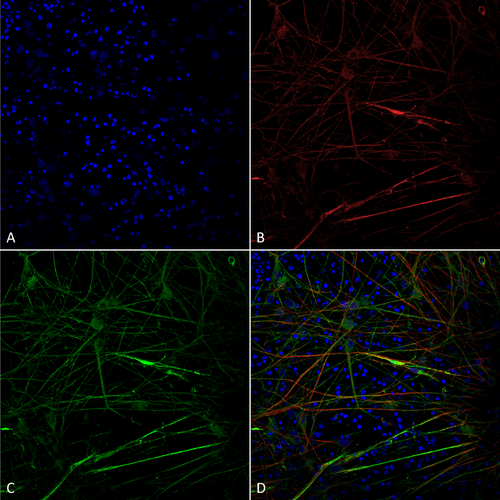
Immunocytochemistry/Immunofluorescence analysis using Mouse Anti-Tau Monoclonal Antibody, Clone 1D5 (SMC-607). Tissue: iPSC-derived neurons. Species: Human. Fixation: 4% PFA. Primary Antibody: Mouse Anti-Tau Monoclonal Antibody (SMC-607) at 1:100 for Overnight at 4°C. Counterstain: DAPI at 1:5000 for 5 minutes at RT in the dark. Magnification: 40X. Courtesy of: Francesco Paonessa.
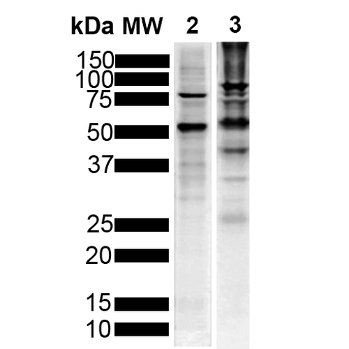
Western Blot analysis of Human, Mouse Breast Cancer Cell line, Brain showing detection of Tau protein using Mouse Anti-Tau Monoclonal Antibody, Clone 1D5 (SMC-607). Lane 1: MW Marker. Lane 2: Human T-47d (10ug). Lane 3: Mouse Brain (20ug).. Block: 5% Skim Milk powder in TBST. Primary Antibody: Mouse Anti-Tau Monoclonal Antibody (SMC-607) at 1:1000 for 2 hours at RT with shaking. Secondary Antibody: Goat anti-mouse IgG:HRP at 1:5000 for 1 hour at RT with shaking. Color Development: Chemiluminescent for HRP (Moss) for 5 min in RT.
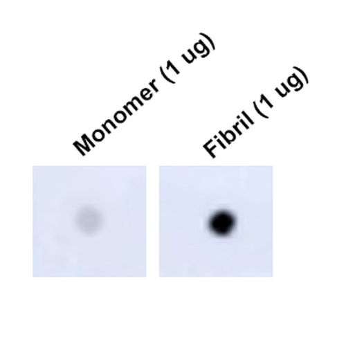
Dot Blot analysis using Mouse Anti-Tau Monoclonal Antibody, Clone 1D5 (SMC-607). Tissue: Recombinant Protein. Species: Human. Primary Antibody: Mouse Anti-Tau Monoclonal Antibody (SMC-607) at 1:1000 for 2 hours at RT with shaking. Secondary Antibody: Goat anti-mouse IgG:HRP at 1:5000 for 1 hour at RT with shaking.
 Powered by Bioz
Reviews
There are no reviews yet.