Discovery through Partnership | Excellence through Quality


Properties
| Storage Buffer | PBS pH7.4, 50% glycerol, 0.09% sodium azide *Storage buffer may change when conjugated |
| Storage Temperature | -20ºC, Conjugated antibodies should be stored according to the product label |
| Shipping Temperature | Blue Ice or 4ºC |
| Purification | Protein G Purified |
| Clonality | Recombinant Monoclonal |
| Clone Number | S74 |
| Isotype | IgG1 |
| Specificity | Detects ~220kDa in extracts of cells transiently transfected with TrpM7. No cross-reactivity against TrpM6. |
| Cite This Product | TRPM7 Antibody (StressMarq Biosciences | Victoria, BC CANADA, Catalog# SMC-316, RRID: AB_2208751) |
| Certificate of Analysis | 1 µg/ml of SMC-316 was sufficient for detection of TrpM7 in 10 µg of COS cell lysate transiently transfected with TprM7 by colorimetric immunoblot analysis using Goat anti-mouse IgG:HRP as the secondary antibody. |
Biological Description
| Alternative Names | CHAK, CHAK1, Channel kinase 1, Channel-kinase 1, Long transient receptor potential channel 7, LTrpC-7, LTRPC7, Transient receptor potential cation channel subfamily M member 7, TRP PLIK, TRPM7, TRPM7_HUMAN |
| Research Areas | ALS Disease, Alzheimer's Disease, Ion Channels, Neurodegeneration, Neuroscience, Parkinson's Disease, Transient Receptor Potential (TRP) Channels |
| Cellular Localization | Membrane |
| Accession Number | NP_001157797.1 |
| Gene ID | 58800 |
| Swiss Prot | Q923J1 |
| Scientific Background |
Transient Receptor Potential Melastatin 7 (TRPM7) is a unique bifunctional protein that combines an ion channel with an α-kinase domain, enabling it to regulate both ionic homeostasis and intracellular signaling. As a divalent cation-permeable channel, TRPM7 is a key regulator of magnesium (Mg²⁺) and calcium (Ca²⁺) influx, essential for neuronal excitability, synaptic plasticity, and cell survival. TRPM7 is widely expressed in the central nervous system and plays a critical role in neuronal development, axonal outgrowth, and glial function. Its kinase activity further modulates cytoskeletal dynamics and gene expression, linking ion transport to broader cellular responses. Dysregulation of TRPM7 has been implicated in several neurodegenerative diseases, including Alzheimer’s disease, Parkinson’s disease, and amyotrophic lateral sclerosis (ALS). In models of ischemia and excitotoxicity, TRPM7 overactivation leads to excessive Ca²⁺ influx, oxidative stress, and neuronal death—hallmarks of neurodegeneration. Moreover, TRPM7-mediated Mg²⁺ dysregulation has been associated with impaired mitochondrial function and neuroinflammation, further contributing to disease progression. Given its dual role in ion transport and kinase signaling, TRPM7 represents a promising therapeutic target in neurodegenerative disease research. Ongoing studies are exploring TRPM7 inhibitors and modulators as potential neuroprotective agents aimed at restoring ionic balance and preventing neuronal loss. |
| References |
1. Brauchi S., Krapivinsky G., Krapivinsky L., Clapham D.E. (2008) Proc Natl Acad Sci USA. 105(24): 8304-8308. 2. Numata T, Okada Y. (2008) J Biol Chem. 283(22): 15097-15103. |
Product Images
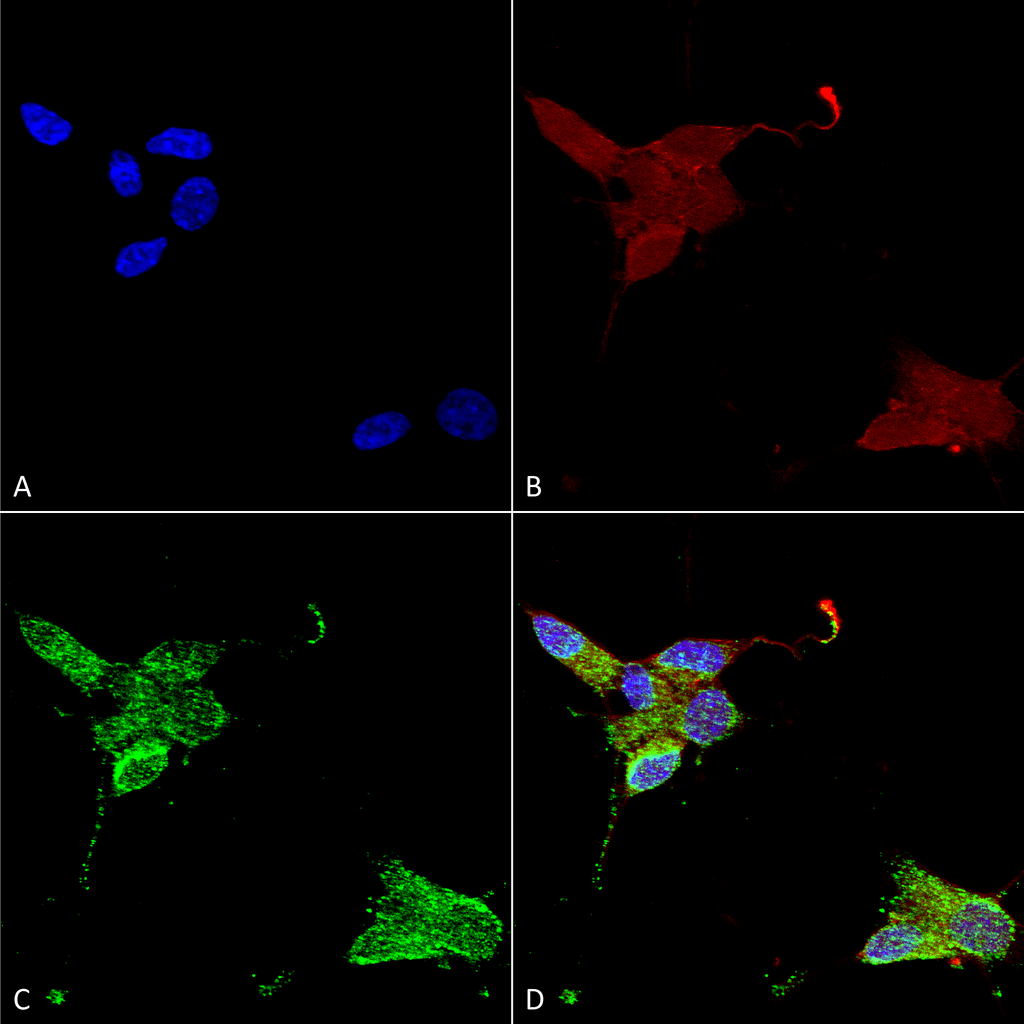
Immunocytochemistry/Immunofluorescence analysis using Mouse Anti-TrpM7 Monoclonal Antibody, Clone S74 (SMC-316). Tissue: Neuroblastoma cells (SH-SY5Y). Species: Human. Fixation: 4% PFA for 15 min. Primary Antibody: Mouse Anti-TrpM7 Monoclonal Antibody (SMC-316) at 1:50 for overnight at 4°C with slow rocking. Secondary Antibody: AlexaFluor 488 at 1:1000 for 1 hour at RT. Counterstain: Phalloidin-iFluor 647 (red) F-Actin stain; Hoechst (blue) nuclear stain at 1:800, 1.6mM for 20 min at RT. (A) Hoechst (blue) nuclear stain. (B) Phalloidin-iFluor 647 (red) F-Actin stain. (C) TrpM7 Antibody (D) Composite.
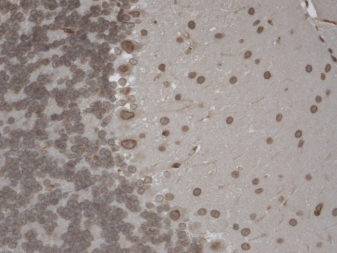
Immunohistochemistry analysis using Mouse Anti-TrpM7 Monoclonal Antibody, Clone S74 (SMC-316). Tissue: Brain Slice. Species: Mouse. Fixation: 10% Formalin Solution for 12-24 hours at RT. Primary Antibody: Mouse Anti-TrpM7 Monoclonal Antibody (SMC-316) at 1:1000 for 1 hour at RT. Secondary Antibody: HRP/DAB Detection System: Biotinylated Goat Anti-Mouse, Streptavidin Peroxidase, DAB Chromogen (brown) for 30 minutes at RT. Counterstain: Mayer Hematoxylin (purple/blue) nuclear stain at 250-500 µl for 5 minutes at RT. Localization: Nuclear staining of both neurons and glia.
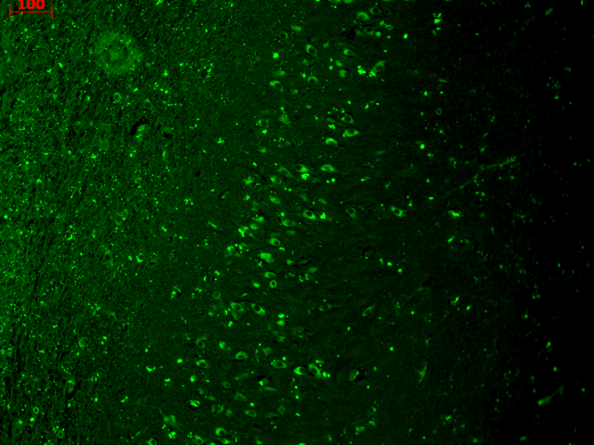
Immunohistochemistry analysis using Mouse Anti-TrpM7 Monoclonal Antibody, Clone S74 (SMC-316). Tissue: hippocampus. Species: Human. Fixation: Bouin’s Fixative and paraffin-embedded. Primary Antibody: Mouse Anti-TrpM7 Monoclonal Antibody (SMC-316) at 1:1000 for 1 hour at RT. Secondary Antibody: FITC Goat Anti-Mouse (green) at 1:50 for 1 hour at RT.
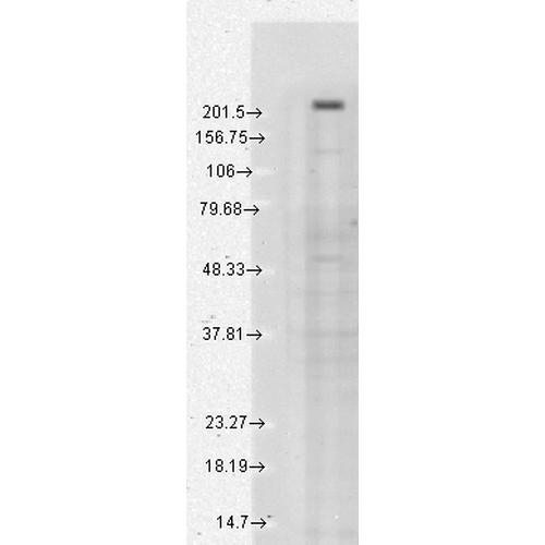
Western Blot analysis of Human cell line mix (A431, A549, HCT116, HeLa, HEK293, HepG2, HL-60, HUVEC, Jurkat, MCF7, PC3, T98G) showing detection of TrpM7 protein using Mouse Anti-TrpM7 Monoclonal Antibody, Clone S74 (SMC-316). Load: 15 µg. Block: 1.5% BSA for 30 minutes at RT. Primary Antibody: Mouse Anti-TrpM7 Monoclonal Antibody (SMC-316) at 1:1000 for 2 hours at RT. Secondary Antibody: Sheep Anti-Mouse IgG: HRP for 1 hour at RT.
 Powered by Bioz
StressMarq Biosciences :
Based on validation through cited publications.