Discovery through Partnership | Excellence through Quality


Properties
| Storage Buffer | PBS pH7.4, 50% glycerol, 0.09% sodium azide *Storage buffer may change when conjugated |
| Storage Temperature | -20ºC, Conjugated antibodies should be stored according to the product label |
| Shipping Temperature | Blue Ice or 4ºC |
| Purification | Protein G Purified |
| Clonality | Monoclonal |
| Clone Number | N15/39 (Formerly sold as S15-39) |
| Isotype | IgG2a |
| Specificity | Detects ~70kDa. |
| Cite This Product | TRPV3 Antibody (StressMarq Biosciences | Victoria, BC CANADA, Catalog# SMC-334, RRID: AB_2209275) |
| Certificate of Analysis | 1 µg/ml of SMC-334 was sufficient for detection of TrpV3 in 10 µg of COS-1 cell lysate transiently transfected with TrpV3 by colorimetric immunoblot analysis using Goat anti-mouse IgG:HRP as the secondary antibody |
Biological Description
| Alternative Names | 1110036I10Rik, MGC124324, MGC124325, Transient receptor potential cation channel subfamily V member 3, TrpV3, Trpv3 heat sensitive channel, TRPV3_HUMAN, Vanilloid receptor 3, Vanilloid receptor like 3, Vanilloid receptor-like 3, vanilloid receptor-related osmotically activated channel protein, VRL 3, VRL-3, VRL3, vanilloid receptor 2, sensitive channel TRPV3 |
| Research Areas | Ion Channels, Neuroscience, Transient Receptor Potential (TRP) Channels |
| Cellular Localization | Membrane |
| Accession Number | NP_001020928 |
| Gene ID | 497948 |
| Swiss Prot | Q4QYD9 |
| Scientific Background |
Transient Receptor Potential Vanilloid 3 (TRPV3) is a non-selective cation channel belonging to the thermosensitive TRP channel family, which plays a critical role in sensory physiology and cellular signaling. Activated by warm temperatures ranging from 22°C to 40°C, TRPV3 is predominantly expressed in keratinocytes and subsets of sensory neurons that innervate the skin. It contributes to temperature sensation, skin barrier function, and vasoregulation. Located on chromosome 17 near the TRPV1 gene, TRPV3 can form heteromeric complexes with other TRP channels, potentially modulating its biophysical properties and expanding its functional repertoire. While traditionally studied in the context of peripheral sensory biology, TRPV3 is increasingly recognized for its relevance in the central nervous system. Emerging evidence suggests that TRPV3 may influence neuroinflammatory pathways, glial activation, and neuronal excitability—processes intimately linked to the pathogenesis of neurodegenerative diseases such as Alzheimer’s and Parkinson’s. Aberrant TRPV3 activity has been associated with altered calcium homeostasis and oxidative stress, both of which are key contributors to neuronal dysfunction and degeneration. As a modulator of both peripheral and central signaling, TRPV3 represents a promising target for therapeutic intervention in neurodegenerative disorders. Ongoing research into its expression patterns, regulatory mechanisms, and interaction networks is shedding new light on its potential role in maintaining neuronal health and resilience. |
| References |
1. Masamoto Y., Kawabata F., Fushiki T. (2009) Biosci. Biotechnol. Biochem. 73(5): 1021-1027. 2. Xiao R., et al. (2008) J Biol Chem. 283(10): 6162-6174. |
Product Images
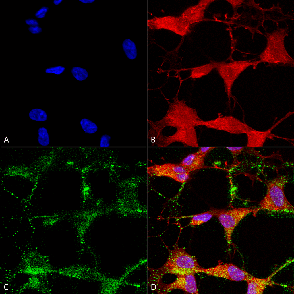
Immunocytochemistry/Immunofluorescence analysis using Mouse Anti-TrpV3 Monoclonal Antibody, Clone S15-39 (SMC-334). Tissue: Neuroblastoma cells (SH-SY5Y). Species: Human. Fixation: 4% PFA for 15 min. Primary Antibody: Mouse Anti-TrpV3 Monoclonal Antibody (SMC-334) at 1:50 for overnight at 4°C with slow rocking. Secondary Antibody: AlexaFluor 488 at 1:1000 for 1 hour at RT. Counterstain: Phalloidin-iFluor 647 (red) F-Actin stain; Hoechst (blue) nuclear stain at 1:800, 1.6mM for 20 min at RT. (A) Hoechst (blue) nuclear stain. (B) Phalloidin-iFluor 647 (red) F-Actin stain. (C) TrpV3 Antibody (D) Composite.
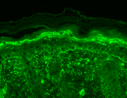
Immunohistochemistry analysis using Mouse Anti-TrpV3 Monoclonal Antibody, Clone S15-39 (SMC-334). Tissue: backskin. Species: Mouse. Fixation: Bouin’s Fixative and paraffin-embedded. Primary Antibody: Mouse Anti-TrpV3 Monoclonal Antibody (SMC-334) at 1:100 for 1 hour at RT. Secondary Antibody: FITC Goat Anti-Mouse (green) at 1:50 for 1 hour at RT. Localization: Filaggrin-like staining in upper layers. Dull lower layer cell staining. Some stain seen in hypodermis.
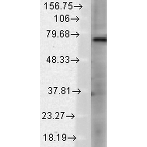
Western Blot analysis of Rat brain membrane lysate showing detection of TrpV3 protein using Mouse Anti-TrpV3 Monoclonal Antibody, Clone S15-39 (SMC-334). Load: 15 µg. Block: 1.5% BSA for 30 minutes at RT. Primary Antibody: Mouse Anti-TrpV3 Monoclonal Antibody (SMC-334) at 1:1000 for 2 hours at RT. Secondary Antibody: Sheep Anti-Mouse IgG: HRP for 1 hour at RT.
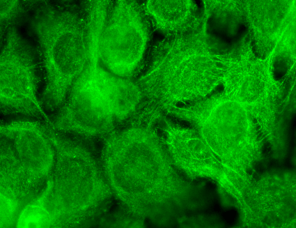
Immunocytochemistry/Immunofluorescence analysis using Mouse Anti-TrpV3 Monoclonal Antibody, Clone S15-39 (SMC-334). Tissue: HaCaT cells. Species: Human. Fixation: Cold 100% methanol for 10 minutes at -20°C. Primary Antibody: Mouse Anti-TrpV3 Monoclonal Antibody (SMC-334) at 1:100 for 1 hour at RT. Secondary Antibody: FITC Goat Anti-Mouse (green) at 1:50 for 1 hour at RT. Localization: Dotty staining in all cells. Some intermediate filament-like staining in some cells.
 Powered by Bioz
StressMarq Biosciences :
Based on validation through cited publications.