Properties
| Storage Buffer | PBS pH7.4, 50% glycerol, 0.1% sodium azide *Storage buffer may change when conjugated |
| Storage Temperature | -20ºC, Conjugated antibodies should be stored according to the product label |
| Shipping Temperature | Blue Ice or 4ºC |
| Purification | Protein G Purified |
| Clonality | Monoclonal |
| Clone Number | N351/23 (Formerly sold as S351-23) |
| Isotype | IgG1 |
| Specificity | Detects >350kDa. |
| Cite This Product | Versican Antibody (StressMarq Biosciences | Victoria, BC CANADA, Catalog# SMC-439, RRID: AB_2701649) |
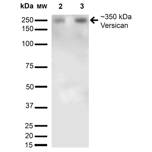
| Certificate of Analysis | 1 µg/ml of SMC-439 was sufficient for detection of Versican in 20 µg of mouse brain membrane lysate and assayed by colorimetric immunoblot analysis using goat anti-mouse IgG:HRP as the secondary antibody. |
Biological Description
| Alternative Names | Versican, CSPG2, VCAN, PG-M, PGM, PG-M proteoglycan, Chondroitin sulfate proteoglycan 2, Chondroitin sulfate proteoglycan core protein 2, Large aggregating proteoglycan, Large fibroblast proteoglycan, Glial hyaluronate-binding protein, Glial hyaluronate binding protein, GHAP, ERVR, Versican core protein, Versican proteoglycan, Versican V0, Versican V1, Versican V2, Versican V3, Versican V4, V1 Neo, WGN, WGN |
| Research Areas | Cell Signaling, Neuroscience, Scaffold Proteins |
| Cellular Localization | Extracellular Matrix, Extracellular Space |
| Accession Number | AAH96495 |
| Gene ID | 13003 |
| Swiss Prot | Q62059 |
| Scientific Background |
Versican (chondroitin sulfate proteoglycan 2, or CSPG2) is a large extracellular matrix (ECM) proteoglycan that plays a pivotal role in neural development, plasticity, and disease. As a key structural component of the brain’s ECM, Versican contributes to the formation of hydrated, viscoelastic matrices that regulate cell proliferation, migration, and differentiation. Its modular structure—featuring hyaluronic acid- and glycosaminoglycan-binding domains, EGF-like repeats, a lectin-like sequence, and a complement regulatory protein-like domain—enables dynamic interactions with neural cells and signaling molecules. Alternative splicing of the VCAN gene produces isoforms with distinct glycosaminoglycan modifications, influencing tissue-specific functions and pathological outcomes. In the central nervous system (CNS), Versican is increasingly recognized for its role in modulating neuroinflammation, glial scar formation, and synaptic remodeling—processes central to neurodegenerative diseases such as Alzheimer’s, Parkinson’s, and multiple sclerosis. Versican-rich pericellular matrices, often co-localized with hyaluronan, can alter cell adhesion and morphology, facilitating neural cell motility and mitosis. These properties are critical in both developmental neurobiology and the maladaptive responses seen in CNS injury and degeneration. Emerging evidence also links Versican dysregulation to impaired neural repair and chronic inflammation, positioning it as a potential biomarker and therapeutic target in neurodegenerative disease research. |
| References |
1. Dours-Zimmermann M.T. and Zimmermann D.R. (1994) J. Biol. Chem. 52: 32992-32998. 2. Evanko S.P., Angello J.C. and Wight T.N. (1999) Arterioscler. Thromb. Vasc. Biol. 4:1004-1013. 3. Lemire J.M., et al. (1999) Arterioscler. Thromb. Vasc. Biol. 7: 1630-1639. 4.Wight T.N. (2002) Curr. Opin. Cell Biol. 5: 617-623. 5. Touab M., Villena J., Barranco C., Arumi-Uria M. and Bassols A. (2002) Am. J. Pathol. 2: 549-557. |
Product Images

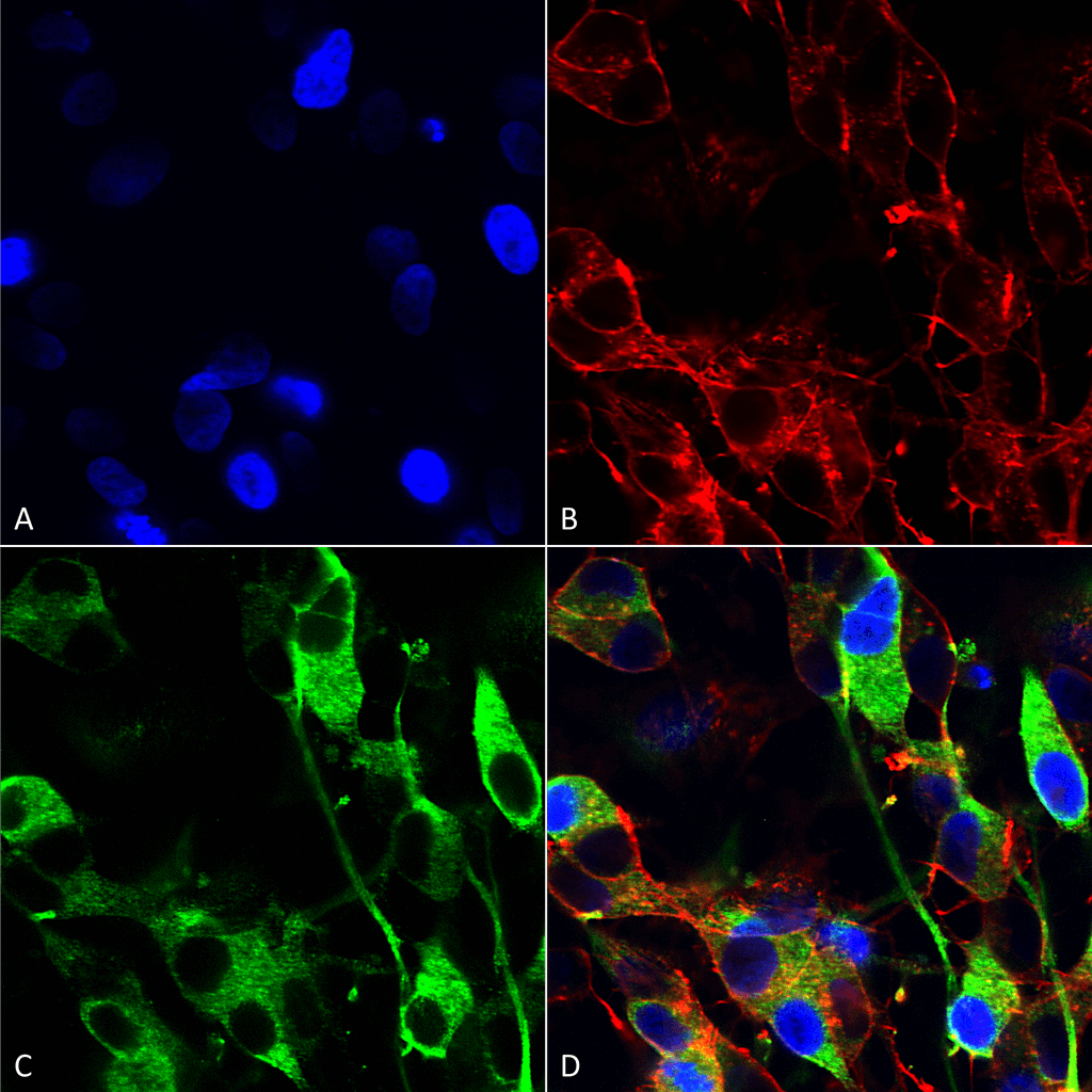
Immunocytochemistry/Immunofluorescence analysis using Mouse Anti-Versican Monoclonal Antibody, Clone N351/23 (SMC-439). Tissue: Neuroblastoma cells (SH-SY5Y). Species: Human. Fixation: 4% PFA for 15 min. Primary Antibody: Mouse Anti-Versican Monoclonal Antibody (SMC-439) at 1:200 for overnight at 4°C with slow rocking. Secondary Antibody: AlexaFluor 488 at 1:1000 for 1 hour at RT. Counterstain: Phalloidin-iFluor 647 (red) F-Actin stain; Hoechst (blue) nuclear stain at 1:800, 1.6mM for 20 min at RT. (A) Hoechst (blue) nuclear stain. (B) Phalloidin-iFluor 647 (red) F-Actin stain. (C) Versican Antibody (D) Composite.

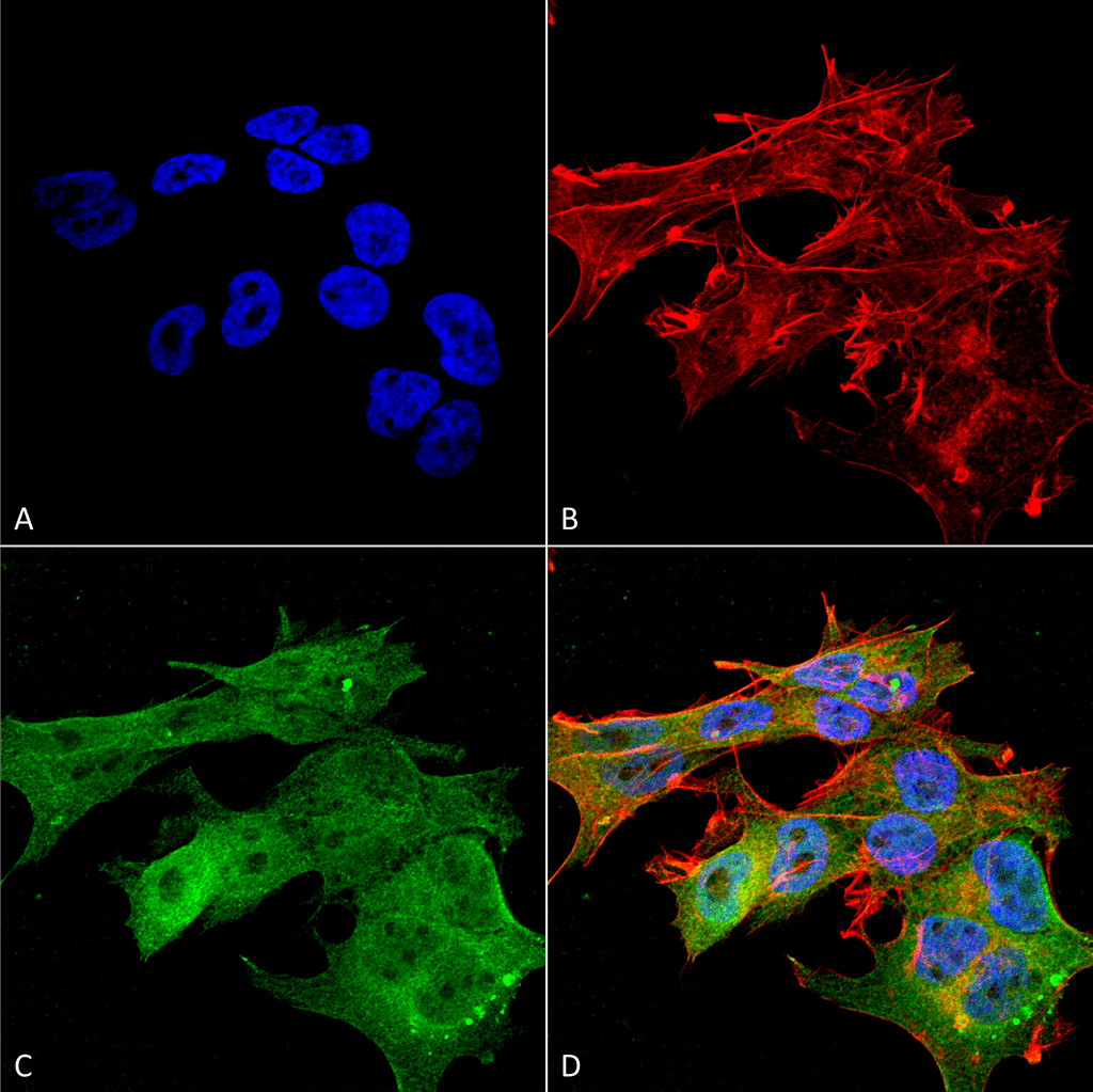
Immunocytochemistry/Immunofluorescence analysis using Mouse Anti-Versican Monoclonal Antibody, Clone N351/23 (SMC-439). Tissue: Neuroblastoma cell line (SK-N-BE). Species: Human. Fixation: 4% Formaldehyde for 15 min at RT. Primary Antibody: Mouse Anti-Versican Monoclonal Antibody (SMC-439) at 1:100 for 60 min at RT. Secondary Antibody: Goat Anti-Mouse ATTO 488 at 1:100 for 60 min at RT. Counterstain: Phalloidin Texas Red F-Actin stain; DAPI (blue) nuclear stain at 1:1000, 1:5000 for 60min RT, 5min RT. Localization: Cytoplasm, Membrane, Extracellular Space, Extracellular Matrix. Magnification: 60X. (A) DAPI (blue) nuclear stain. (B) Phalloidin Texas Red F-Actin stain. (C) Versican Antibody. (D) Composite.

Western Blot analysis of Rat Brain Membrane and brain showing detection of 350kDa Versican protein using Mouse Anti-Versican Monoclonal Antibody, Clone N351/23 (SMC-439). Lane 1: Molecular Weight Ladder. Lane 2: Rat Brain Membrane. Lane 3: Rat Brain. Load: 15 µg. Block: 5% Skim Milk in 1X TBST. Primary Antibody: Mouse Anti-Versican Monoclonal Antibody (SMC-439) at 1:200 for 16 hours at 4°C. Secondary Antibody: Goat Anti-Mouse IgG: HRP at 1:1000 for 1 hour RT. Color Development: KPL TMB. Predicted/Observed Size: 350kDa. Other Band(s): Multiple other bands.


Reviews
There are no reviews yet.