Discovery through Partnership | Excellence through Quality


Properties
| Storage Buffer | PBS pH7.4, 50% glycerol, 0.09% sodium azide *Storage buffer may change when conjugated |
| Storage Temperature | -20ºC, Conjugated antibodies should be stored according to the product label |
| Shipping Temperature | Blue Ice or 4ºC |
| Purification | Protein G Purified |
| Clonality | Monoclonal |
| Clone Number | N28/9 (Formerly sold as S28-9) |
| Isotype | IgG1 |
| Specificity | Detects ~52kDa. No cross-reactivity against VGlut2. |
| Cite This Product | VGLUT1 Antibody (StressMarq Biosciences | Victoria, BC CANADA, Catalog# SMC-394, RRID: AB_11229710) |
| Certificate of Analysis | 1 µg/ml of SMC-394 was sufficient for detection of VGLut1 in 20 µg of rat brain lysate by colorimetric immunoblot analysis using goat anti-mouse IgG:HRP as the secondary antibody. |
Biological Description
| Alternative Names | VGLUT1, Vesicular Glutamate Transporter 1, SLC17A7, BNPI, Solute Carrier Family 17 Member 7, Brain-specific Na(+)-dependent Inorganic Phosphate Cotransporter, Solute Carrier Family 17 (Sodium-Dependent Inorganic Phosphate Cotransporter), Member 7, Solute Carrier Family 17 (Vesicular Glutamate Transporter), Member 7, VGluT1, VGLUT 1 |
| Research Areas | Cell Markers, Cell Signaling, Neuron Markers, Neuroscience, Neurotransmitter Transporters, Presynaptic Markers, Pumps/Transporters |
| Cellular Localization | Cell Junction, Cytoplasmic Vesicle, Membrane, Secretory vesicle, Synapse, Synaptic vesicle membrane |
| Accession Number | NP_446311.1 |
| Gene ID | 116638 |
| Swiss Prot | Q62634 |
| Scientific Background |
Vesicular glutamate transporter 1 (VGLUT1) plays a critical role in glutamatergic neurotransmission by packaging glutamate into synaptic vesicles for release at excitatory synapses. Expressed in a specific subset of glutamatergic neurons, VGLUT1 exhibits a unique chloride conductance that is inhibited by glutamate, highlighting its dual role in vesicle loading and ion homeostasis. Unlike high-affinity plasma membrane excitatory amino acid transporters (EAATs), VGLUT1 operates with a lower apparent affinity, with a Km of approximately 2 mM—comparable to native synaptic vesicle transport kinetics. Importantly, VGLUT1 demonstrates substrate specificity by selectively transporting glutamate, but not aspartate, distinguishing it from EAATs that recognize both. This specificity underscores VGLUT1’s essential function in maintaining synaptic fidelity and excitatory signaling precision. Emerging evidence links dysregulation of VGLUT1 expression and function to several neurodegenerative disorders, including Alzheimer’s disease, Parkinson’s disease, and amyotrophic lateral sclerosis (ALS). Altered glutamate homeostasis, driven by impaired vesicular transport, contributes to excitotoxicity—a key pathological mechanism in these conditions. As a result, VGLUT1 has become a focal point in neuroscience and neurodegenerative disease research, offering potential as both a biomarker and a therapeutic target. Understanding its molecular properties and regulatory mechanisms is essential for developing strategies to modulate glutamatergic signaling in disease contexts. |
| References |
1. Wojcik S.M., et al. (2004) PNAS. 101(18): 7158-7163. 2. Shigeri Y., Seal R.P., Shimamoto K. (2004) Brain Res Rev. 45(3): 250-265. |
Product Images
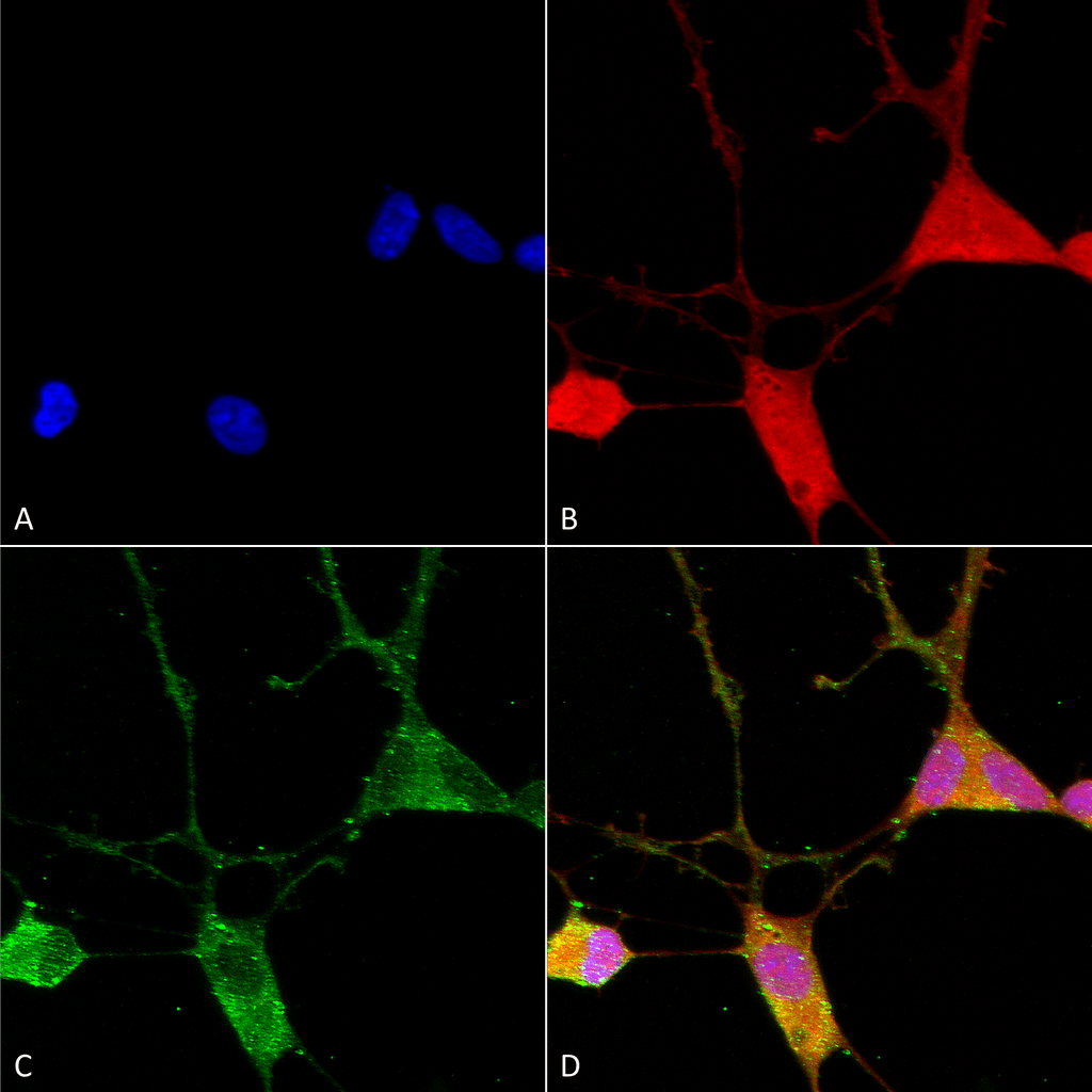
Immunocytochemistry/Immunofluorescence analysis using Mouse Anti-VGLUT1 Monoclonal Antibody, Clone N28/9 (SMC-394). Tissue: Neuroblastoma cells (SH-SY5Y). Species: Human. Fixation: 4% PFA for 15 min. Primary Antibody: Mouse Anti-VGLUT1 Monoclonal Antibody (SMC-394) at 1:100 for overnight at 4°C with slow rocking. Secondary Antibody: AlexaFluor 488 at 1:1000 for 1 hour at RT. Counterstain: Phalloidin-iFluor 647 (red) F-Actin stain; Hoechst (blue) nuclear stain at 1:800, 1.6mM for 20 min at RT. (A) Hoechst (blue) nuclear stain. (B) Phalloidin-iFluor 647 (red) F-Actin stain. (C) VGLUT1 Antibody (D) Composite.
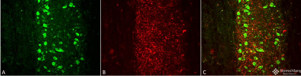
Immunohistochemistry analysis using Mouse Anti-VGLUT1 Monoclonal Antibody, Clone N28/9 (SMC-394). Tissue: spinal cord. Species: Mouse. Fixation: 4% PFA. Primary Antibody: Mouse Anti-VGLUT1 Monoclonal Antibody (SMC-394) at 1:500 for 16 hours at RT. Secondary Antibody: Alexa Fluor 555 Donkey Anti-Mouse (red) at 1:2000 for 2 hours at RT. Counterstain: NeuN neuronal stain (green). Magnification: 20X. Courtesy of: Leilei Wang, Ph.D. UT Southwestern Medical Center at Dallas.
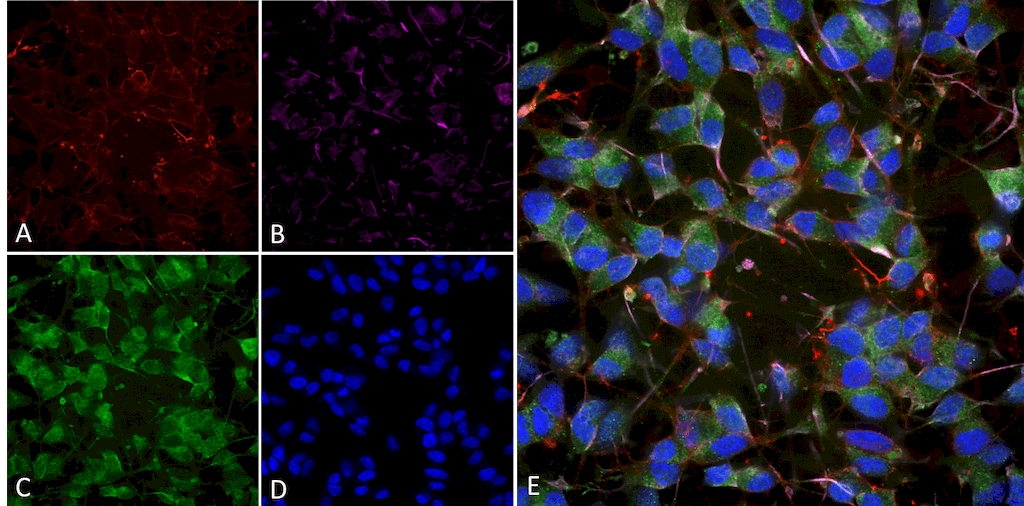
Immunocytochemistry/Immunofluorescence analysis using Mouse Anti-VGLUT1 Monoclonal Antibody, Clone N28/9 (SMC-394). Tissue: Differentiated SH-SY5Y. Species: Human. Primary Antibody: Mouse Anti-VGLUT1 Monoclonal Antibody (SMC-394) at 1:100. Secondary Antibody: AlexaFluor 488. Counterstain: phalloidin (Alexa 647, red), beta tubulin (Anti-beta III Tubulin Ab, Alexa 555, magenta) Hoechst (blue). (A) Phalloidin (B) Anti-beta III Tubulin Ab. (C) VGLUT1 Antibody. (D) Hoechst (E) Composite.
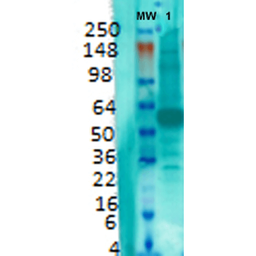
Western Blot analysis of Rat brain membrane lysate showing detection of VGLUT1 protein using Mouse Anti-VGLUT1 Monoclonal Antibody, Clone N28/9 (SMC-394). Primary Antibody: Mouse Anti-VGLUT1 Monoclonal Antibody (SMC-394) at 1:1000.
 Powered by Bioz
StressMarq Biosciences :
Based on validation through cited publications.