Discovery through Partnership | Excellence through Quality


Properties
| Storage Buffer | PBS pH 7.4, 50% glycerol, 0.09% Sodium azide *Storage buffer may change when conjugated |
| Storage Temperature | -20ºC, Conjugated antibodies should be stored according to the product label |
| Shipping Temperature | Blue Ice or 4ºC |
| Purification | Protein G Purified |
| Clonality | Monoclonal |
| Clone Number | 10A8 |
| Isotype | IgG2a |
| Specificity | Detects ~92 kDa. |
| Cite This Product | VPS35 Antibody (StressMarq Biosciences | Victoria, BC CANADA, Catalog# SMC-605, RRID: AB_2820304) |
| Certificate of Analysis | A 1:1000 dilution of SMC-605 was sufficient for detection of VPS35 in 10 µg of SH-SY5Y by ECL immunoblot analysis using Goat Anti-Mouse IgG:HRP as the secondary antibody. |
Biological Description
| Alternative Names | VPS35, VPS35 Retromer Complex Component, Vacuolar Protein Sorting-Associated Protein 35, Vacuolar Protein Sorting 35 Homolog, Vesicle Protein Sorting 35, MEM3, PARK17, FLJ10752, HVPS35, Maternal-Embryonic 3, TCCCTA00141 |
| Research Areas | Alzheimer's Disease, Cell Signaling, Golgi Proteins, Membrane Trafficking Proteins, Neurodegeneration, Neuroscience, Parkinson's Disease, Protein Trafficking |
| Cellular Localization | Cytoplasm, Endosome, Lysosome, Membrane, Vesicles |
| Accession Number | NP_060676.2 |
| Gene ID | 55737 |
| Swiss Prot | Q96QK1 |
| Scientific Background |
Vacuolar Protein Sorter 35 (VPS35) is a core component of the retromer complex, a critical intracellular trafficking system responsible for the endosome-to-Golgi retrieval of membrane proteins. This pathway is essential for maintaining protein homeostasis, synaptic function, and neuronal survival. Mutations in VPS35—most notably the D620N variant—have been directly linked to familial and sporadic forms of Parkinson’s disease (PD). These mutations impair retromer function, leading to disrupted recycling of key neuronal receptors and transporters. Dysfunctional VPS35 also contributes to mitochondrial fragmentation, oxidative stress, and impaired autophagy—cellular processes that are central to the pathogenesis of neurodegenerative diseases. Beyond Parkinson’s, VPS35 dysfunction has been implicated in Alzheimer’s disease and other proteinopathies, where it affects the trafficking of amyloid precursor protein (APP) and tau. Its role in regulating endosomal sorting and mitochondrial dynamics positions VPS35 as a critical node in the cellular pathways that maintain neuronal integrity. As a result, VPS35 is emerging as a promising therapeutic target in neurodegenerative disease research. Restoring or enhancing retromer function may offer novel strategies to counteract protein misfolding, synaptic loss, and neuroinflammation. |
| References |
1. Vilarino-Guell, C. et al. (2011) Am J Hum Genet 89:162–167 2. Zimprich, A. et al. (2011) Am J Hum Genet 89:168–175 3. Rahman, A.A., Morrison, B.E. (2019) Neurosci 401:1-10. |
Product Images
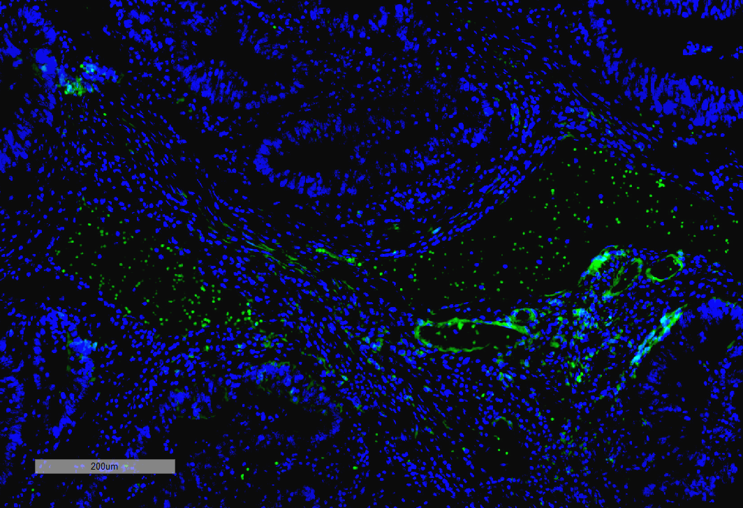
Immunohistochemistry analysis using Mouse Anti-VPS35 Monoclonal Antibody, Clone 10A8 (SMC-605). Tissue: Thyroid Cancer. Species: Human. Primary Antibody: Mouse Anti-VPS35 Monoclonal Antibody (SMC-605) at 1:100 for Overnight at 4C, then 30 min at 37C. Secondary Antibody: Goat Anti-Mouse IgG (H+L): FITC for 45 min at 37C. Counterstain: DAPI for 3 min at RT. Magnification: 10X.
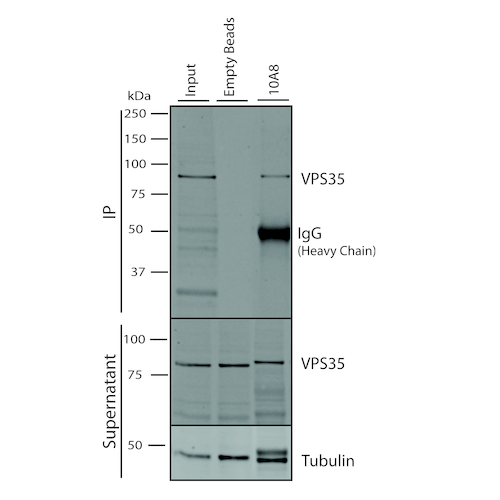
Immunoprecipitation analysis using Mouse Anti-VPS35 Monoclonal Antibody, Clone 10A8 (SMC-605). Tissue: A549 cells. Species: Human. Primary Antibody: Mouse Anti-VPS35 Monoclonal Antibody (SMC-605). 500 µL cell culture supernatants were incubated with 10 µL of Protein A/G resin beads for 1 hour at 4⁰C. SMC-605 clone 10A8 depletes VPS35 from the A549 cell extract..
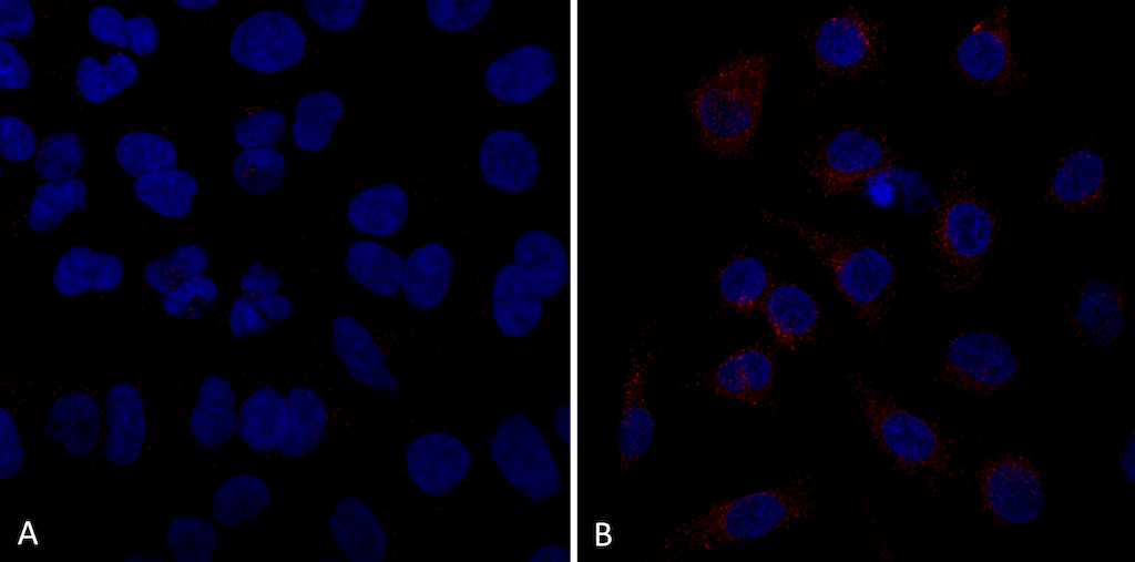
Immunocytochemistry/Immunofluorescence analysis using Mouse Anti-VPS35 Monoclonal Antibody, Clone 10A8 (SMC-605). Tissue: A549 cells. Species: Human. Primary Antibody: Mouse Anti-VPS35 Monoclonal Antibody (SMC-605) at 1:5 (tissue culture supernatant). Secondary Antibody: Donkey anti-mouse: Alexa Fluor 594 at 1:4000 in 0.2% BSA PBS. Counterstain: DAPI. Localization: Vesicles. A) VPS35 KO A549 cells B) WT A549 cells. Courtesy of: Dario Alessi Lab, University of Dundee.
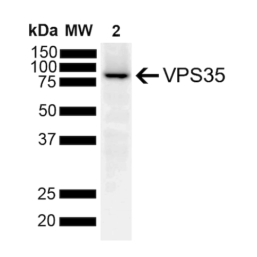
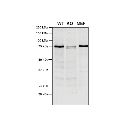
Western Blot analysis of Human, Mouse A549, MEF showing detection of VPS35 protein using Mouse Anti-VPS35 Monoclonal Antibody, Clone 10A8 (SMC-605). Lane 1: Molecular Weight Ladder. Lane 2: VPS35 KO A549 cells. Lane 3: mouse embryonic fibroblast cells.. Load: 8 µg each A549 and MEF. Primary Antibody: Mouse Anti-VPS35 Monoclonal Antibody (SMC-605) at 1:5 (tissue culture supernatant). Secondary Antibody: Donkey anti-mouse IRDye 800CW at 1:25000 in TBS-T.
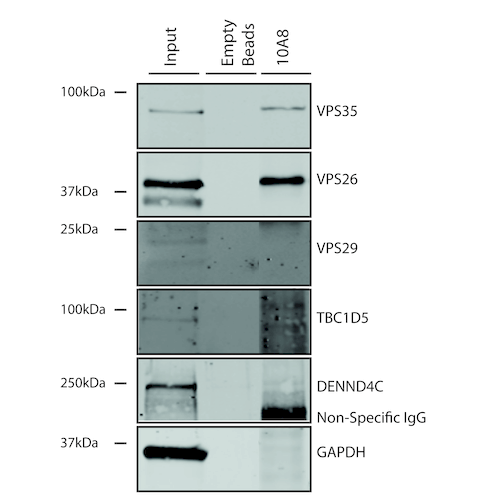
Immunoprecipitation analysis using Mouse Anti-VPS35 Monoclonal Antibody, Clone 10A8 (SMC-605). Tissue: A549 cells. Species: Human. Primary Antibody: Mouse Anti-VPS35 Monoclonal Antibody (SMC-605).
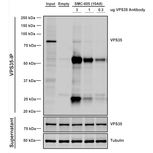
Immunoprecipitation analysis using Mouse Anti-VPS35 Monoclonal Antibody, Clone 10A8 (SMC-605). Tissue: A549 cells. Species: Human. Primary Antibody: Mouse Anti-VPS35 Monoclonal Antibody (SMC-605). Three amounts of SMC-605 (3, 1 and 0.3 ug) were non-covalently coupled to 10uL of A/G sepharose beads for 1 hour at 4°C and next incubated with 250ug of A549 lysate for 2 hours at 4°C.
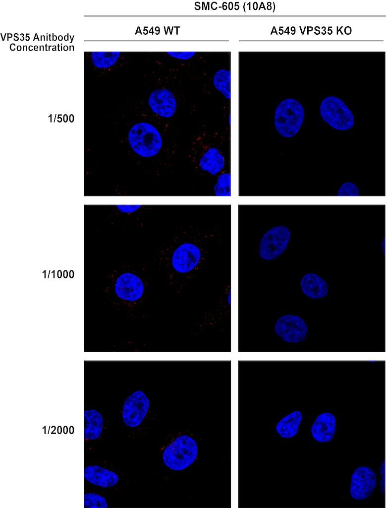
Immunocytochemistry/Immunofluorescence analysis using Mouse Anti-VPS35 Monoclonal Antibody, Clone 10A8 (SMC-605). Tissue: A549 WT, VPS35 KO cells. Species: Human. Primary Antibody: Mouse Anti-VPS35 Monoclonal Antibody (SMC-605). Secondary Antibody: Donkey Anti-Mouse AlexaFluor 594. Clone can detect VPS35 at 1/2000 concentration.
 Powered by Bioz
StressMarq Biosciences :
Based on validation through cited publications.