Discovery through Partnership | Excellence through Quality


Properties
| Storage Buffer | PBS pH7.4, 50% glycerol, 0.09% sodium azide *Storage buffer may change when conjugated |
| Storage Temperature | -20ºC, Conjugated antibodies should be stored according to the product label |
| Shipping Temperature | Blue Ice or 4ºC |
| Purification | Affinity Purified |
| Clonality | Polyclonal |
| Specificity | Detects ~22kDa. Does not cross-react with αA-crystallin. |
| Cite This Product | Alpha B Crystallin Antibody (StressMarq Biosciences | Victoria, BC CANADA, Catalog# SPC-126, RRID: AB_2084306) |
| Certificate of Analysis | A 1:5000 dilution of SPC-126 was sufficient for detection of alpha B crystallin in 20 µg of HeLa cell lysate by ECL immunoblot analysis. |
Biological Description
| Alternative Names | Alpha B Crystallin, CRYAB, CRYA2, HSPB5, Heat Shock Protein Beta-5, Heat Shock Protein Family B Member 5, Alpha-Crystallin B Chain, Crystallin Alpha B, Alpha (B)-Crystallin, HEL-S-101, CMD1II, CTPP2, CTRCT16, MFM2, Rosenthal Fiber Component, Epididymis Secretory Protein Li 101, Heat-Shock 20 KD Like-Protein, Renal Carcinoma Antigen NY-REN-27 |
| Research Areas | Cancer, Cell Signaling, Chaperone Proteins, Heat Shock, Neuroscience, Protein Trafficking |
| Cellular Localization | Cytoplasm, Nucleus |
| Accession Number | NP_001876.1 |
| Gene ID | 1410 |
| Swiss Prot | P02511 |
| Scientific Background |
Alpha B Crystallin (CRYAB) is a small heat shock protein (sHSP) originally identified as a structural component of the vertebrate eye lens. While it contributes to lens transparency by preventing protein aggregation, CRYAB is now recognized for its broader role as a molecular chaperone and cytoprotective agent in various tissues, including the central nervous system. CRYAB shares structural and functional similarities with other sHSPs such as HSP25 and HSP27. It binds to misfolded or denatured proteins, stabilizing them in a soluble state and preventing toxic aggregation—a key pathological feature in neurodegenerative diseases like Alzheimer’s, Parkinson’s, and multiple sclerosis. Under cellular stress, CRYAB becomes phosphorylated, enhancing its chaperone activity and contributing to protein quality control. Unlike its Alpha A counterpart, Alpha B Crystallin is widely expressed beyond the lens, including in glial cells, neurons, and muscle tissue. It interacts with diverse cellular components such as membrane proteins, cytoskeletal elements, nuclear proteins, and DNA, supporting its role in maintaining intracellular architecture and stress resilience. Importantly, CRYAB is overexpressed in several neurological disorders and has been shown to inhibit apoptosis by blocking caspase activation. This anti-apoptotic function positions CRYAB as a promising therapeutic target and biomarker in neurodegenerative disease research, with potential applications in neuroprotection, inflammation modulation, and regenerative medicine. |
| References |
1. Merck K.B. et al. (1993) J Biol Chem. 268: 1046-1052. 2. Horwitz J. (1992) Proc Natl Acad Sci USA 89(21): 10449-10453. 3. Cobb B.A. and Petrash J.M. (2002) Biochemistry. 41: 483-490 4. Horwitz J. (2003) Exp Eye Res. 76: 145-153. 5. Bullard B. et al. (2004) J Biol Chem. 279: 7917-7924. 6. Gangalum R.K., Schibler M.J. and Bhat S.P. (2004) J Biol Chem. 279: 43374.43377. 7. Maddala R. and Rao V.P. (2005) Exp Cell Res. 306: 203-215. 8. Yaung J., et al. (2007) Molecular Vision 13: 566-577. 9. Head M.W. et al. (2000) Neuropathol Appl Neurobiol. 26: 304-312. |
Product Images
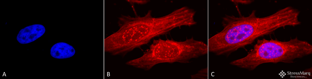
Immunocytochemistry/Immunofluorescence analysis using Rabbit Anti-Alpha B Crystallin Polyclonal Antibody (SPC-126). Tissue: Heat Shocked Cervical cancer cell line (HeLa). Species: Human. Fixation: 2% Formaldehyde for 20 min at RT. Primary Antibody: Rabbit Anti-Alpha B Crystallin Polyclonal Antibody (SPC-126) at 1:120 for 12 hours at 4°C. Secondary Antibody: APC Goat Anti-Rabbit (red) at 1:200 for 2 hours at RT. Counterstain: DAPI (blue) nuclear stain at 1:40000 for 2 hours at RT. Localization: Actin filament bundles. Nuclear splicing speckles. Exosomes. Magnification: 100x. (A) DAPI (blue) nuclear stain. (B) Anti-Alpha B Crystallin Antibody. (C) Composite. Heat Shocked at 42°C for 1h.
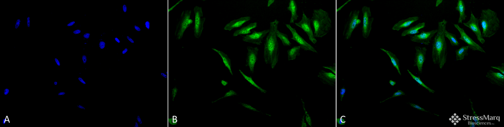
Immunocytochemistry/Immunofluorescence analysis using Rabbit Anti-Alpha B Crystallin Polyclonal Antibody (SPC-126). Tissue: Heat Shocked Cervical cancer cell line (HeLa). Species: Human. Fixation: 2% Formaldehyde for 20 min at RT. Primary Antibody: Rabbit Anti-Alpha B Crystallin Polyclonal Antibody (SPC-126) at 1:120 for 12 hours at 4°C. Secondary Antibody: FITC Goat Anti-Rabbit (green) at 1:200 for 2 hours at RT. Counterstain: DAPI (blue) nuclear stain at 1:40000 for 2 hours at RT. Localization: Actin filament bundles. Nuclear splicing speckles. Exosomes. Magnification: 20x. (A) DAPI (blue) nuclear stain. (B) Anti-Alpha B Crystallin Antibody. (C) Composite. Heat Shocked at 42°C for 1h.
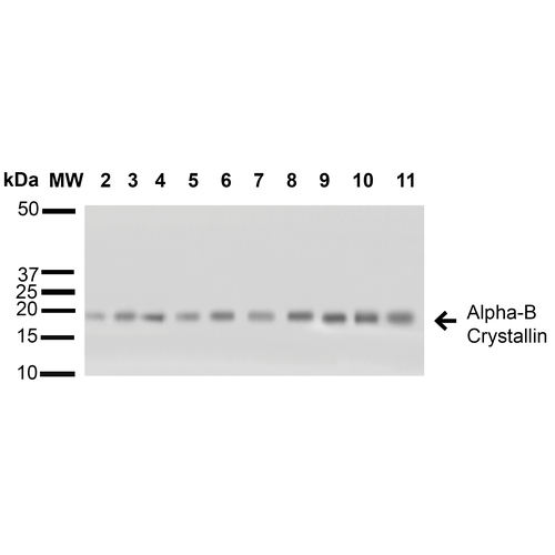
Western blot analysis of Human A431, HCT116, HeLa, HepG2, HEK293, HUVEC, Jurkat, MCF7, PC3 and T98G cell lysates showing detection of ~22 kDa Alpha B Crystallin protein using Rabbit Anti-Alpha B Crystallin Polyclonal Antibody (SPC-126). Lane 1: Molecular Weight Ladder (MW). Lane 2: A431 cell lysates. Lane 3: HCT116 cell lysates. Lane 4: HeLa cell lysates. Lane 5: HepG2 cell lysates. Lane 6: HEK293 cell lysates. Lane 7: HUVEC cell lysates. Lane 8: Jurkat cell lysates. Lane 9: MCF7 cell lysates. Lane 10: PC3 cell lysates. Lane 11: T98G cell lysates. Load: 15 µg. Block: 5% Skim Milk in 1X TBST. Primary Antibody: Rabbit Anti-Alpha B Crystallin Polyclonal Antibody (SPC-126) at 1:1000 for 60 min at RT. Secondary Antibody: Goat Anti-Rabbit IgG: HRP at 1:1000 for 60 min at RT. Color Development: ECL solution for 6 min in RT. Predicted/Observed Size: ~22 kDa.
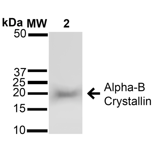
Western blot analysis of Rat Brain cell lysates showing detection of ~22 kDa Alpha B Crystallin protein using Rabbit Anti-Alpha B Crystallin Polyclonal Antibody (SPC-126). Lane 1: Molecular Weight Ladder (MW). Lane 2: Rat Brain cell lysates. Load: 15 µg. Block: 5% Skim Milk in 1X TBST. Primary Antibody: Rabbit Anti-Alpha B Crystallin Polyclonal Antibody (SPC-126) at 1:1000 for 60 min at RT. Secondary Antibody: Goat Anti-Rabbit IgG: HRP at 1:1000 for 60 min at RT. Color Development: ECL solution for 6 min in RT. Predicted/Observed Size: ~22 kDa.
 Powered by Bioz
StressMarq Biosciences :
Based on validation through cited publications.