Discovery through Partnership | Excellence through Quality


Properties
| Storage Buffer | PBS, 50% glycerol, 0.09% sodium azide *Storage buffer may change when conjugated |
| Storage Temperature | -20ºC, Conjugated antibodies should be stored according to the product label |
| Shipping Temperature | Blue Ice or 4ºC |
| Purification | Protein A purified |
| Clonality | Polyclonal |
| Specificity | Recognizes generic epitopes common to many amyloid fibrils and fibrillar oligomers, but not prefibrillar oligomers or natively folded proteins. Expected to detect in Mouse and Rat based on species homology. |
| Cite This Product | Amyloid Fibrils (OC) Antibody (StressMarq Biosciences | Victoria, BC CANADA, Catalog# SPC-507, RRID: AB_10960639) |
| Certificate of Analysis | A 1:1000 dilution of SPC-507 was sufficient for detection of amyloid fibrils on PVDF membranes using transferred fibrils by colorimetric dot blot analysis using Goat anti-rabbit IgG:HRP as the secondary antibody. |
Biological Description
| Alternative Names | OC, Fibrils, Amyloid Oligomer aß, A11, Amyloid beta A4 protein, ABPP, APPI, Alzheimer disease amyloid protein, Cerebral vascular amyloid peptide, PreA4, Protease nexin-II, APP, A4, AD, Amyloid precursor protein, Amyloid-β |
| Research Areas | Alzheimer's Disease, Amyloid, Blood, Cardiovascular System, Cell Signaling, Neurodegeneration, Neuroscience |
| Cellular Localization | Membrane |
| Scientific Background |
Amyloid fibrils are highly ordered protein aggregates formed through the misfolding and oligomerization of normally soluble proteins. These fibrillar structures, often rich in β-sheet content, are hallmarks of numerous neurodegenerative diseases (1,2). Even non-disease-related proteins can adopt amyloidogenic conformations under conditions of partial unfolding or denaturation, leading to the formation of toxic aggregates. In neurodegenerative research, amyloid fibrils—particularly those recognized by the OC antibody, which detects fibrillar oligomers—are critical biomarkers of disease progression and pathology. Their accumulation disrupts cellular homeostasis, impairs synaptic function, and triggers neuroinflammation. Prominent examples include amyloid-β (Aβ) plaques and tau neurofibrillary tangles in Alzheimer’s disease, α-synuclein aggregates in the Lewy bodies of Parkinson’s disease, and polyglutamine-rich inclusions in Huntington’s disease (2,3). These fibrillar assemblies are not merely byproducts but active contributors to neuronal dysfunction and cell death. Understanding the structural and biochemical properties of amyloid fibrils is essential for developing targeted diagnostics and therapeutics. The OC antibody, which selectively binds to fibrillar but not prefibrillar or monomeric species, has become a valuable tool in distinguishing toxic conformers and mapping disease-specific aggregation pathways. As research advances, amyloid fibrils remain at the forefront of neurodegenerative disease studies, offering insights into protein misfolding disorders and potential avenues for intervention. |
| References |
1. Glabe C.G. (2004) Trends Biochem Sci. 29(10): 542-547. 2. Kayed R., et al. (2004) J Bio. Chem. 279: 46363-46366. 3. Kayed R., et al. (2003) Science. 300(5618): 486-489. |
Product Images
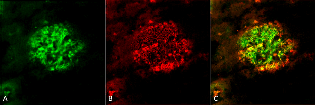
Immunohistochemistry analysis using Rabbit Anti-Amyloid Fibrils (OC) Polyclonal Antibody (SPC-507). Tissue: Alzheimer’s Disease brain. Species: Human. Fixation: Formalin fixed. Primary Antibody: Rabbit Anti-Amyloid Fibrils (OC) Polyclonal Antibody (SPC-507) at 1:5000. Secondary Antibody: Goat Anti-Rabbit ATTO 488 (green). Localization: Plaque. (A) Amyloid Fibril (OC) Antibody (SPC-507). (B) Amyloid Oligomer (A11) Antibody (SPC-506). (C) Composite. Courtesy of: Dr. Elizabeth Head, University of California, Irvine.
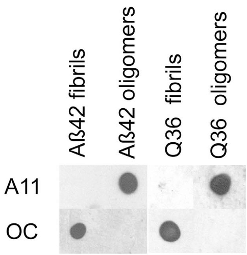
Dot blot analysis using Rabbit Anti-Amyloid Fibrils (OC) Polyclonal Antibody (SPC-507). Tissue: Abeta42 fibrils and prefibrillar oligomers. Species: Human. Primary Antibody: Rabbit Anti-Amyloid Fibrils (OC) Polyclonal Antibody (SPC-507) at 1:1000. Courtesy of: Kayed, R., Head, E., Thompson, J. L., McIntire, T. M., Milton, S. C., Cotman, C. W., et al. (2003). Common structure of soluble amyloid oligomers implies common mechanism of pathogenesis. Science 300, 486–489. doi: 10.1126/science.1079469.
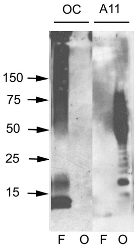
Western blot analysis of Human Abeta42 fibrils and prefibrillar oligomers showing detection of Amyloid Fibrils (OC) protein using Rabbit Anti-Amyloid Fibrils (OC) Polyclonal Antibody (SPC-507). Primary Antibody: Rabbit Anti-Amyloid Fibrils (OC) Polyclonal Antibody (SPC-507) at 1:1000. Courtesy of: Kayed, R., Head, E., Thompson, J. L., McIntire, T. M., Milton, S. C., Cotman, C. W., et al. (2003). Common structure of soluble amyloid oligomers implies common mechanism of pathogenesis. Science 300, 486–489. doi: 10.1126/science.1079469.
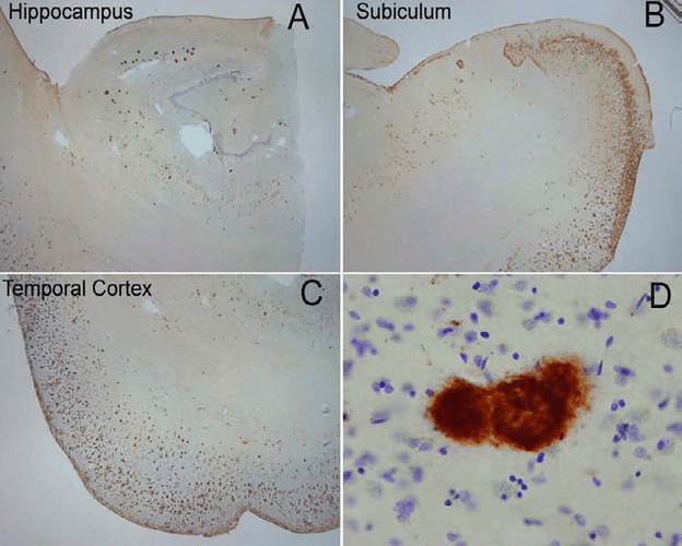
Immunohistochemistry analysis using Rabbit Anti-Amyloid Fibrils (OC) Polyclonal Antibody (SPC-507). Tissue: Alzheimer’s Disease brain. Species: Human. Primary Antibody: Rabbit Anti-Amyloid Fibrils (OC) Polyclonal Antibody (SPC-507) at 1:100. Extensive OC labeling was observed in the hippocampus (A), subiculum (B) and frontal cortex (C) in Alzheimer disease. A higher magnification image illustrates that OC positive deposits were dense and consisted of fine fibrillar material (D). Courtesy of: Kayed, R., Head, E., Thompson, J. L., McIntire, T. M., Milton, S. C., Cotman, C. W., et al. (2003). Common structure of soluble amyloid oligomers implies common mechanism of pathogenesis. Science 300, 486–489. doi: 10.1126/science.1079469.
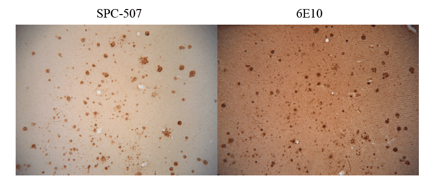
Immunohistochemistry analysis using Rabbit Anti-Amyloid Fibrils (OC) Polyclonal Antibody (SPC-507). Tissue: Alzheimer’s Disease brain. Species: Human. Primary Antibody: Rabbit Anti-Amyloid Fibrils (OC) Polyclonal Antibody (SPC-507) at 1:100. Showing no Amyloid Precursor Protein (APP) cross-reactivity (L), but when conducted with monoclonal 6E10 (R) shows considerable APP cross-reactivity. Courtesy of: Kayed, R., Head, E., Thompson, J. L., McIntire, T. M., Milton, S. C., Cotman, C. W., et al. (2003). Common structure of soluble amyloid oligomers implies common mechanism of pathogenesis. Science 300, 486–489. doi: 10.1126/science.1079469.
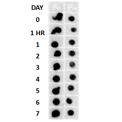
Dot blot analysis using Rabbit Anti-Amyloid Fibrils (OC) Polyclonal Antibody (SPC-507). Tissue: Cell lysates. Species: Human. Primary Antibody: Rabbit Anti-Amyloid Fibrils (OC) Polyclonal Antibody (SPC-507) at 1:500, 1:5000. Beta Amyloid HEPES-NaCl aggregation, showing 1:500 (L) and 1:5000 (R) time lapse dot blot.
 Powered by Bioz
StressMarq Biosciences :
Based on validation through cited publications.