Discovery through Partnership | Excellence through Quality


Properties
| Storage Buffer | PBS, 50% glycerol, 0.09% sodium azide *Storage buffer may change when conjugated |
| Storage Temperature | -20ºC, Conjugated antibodies should be stored according to the product label |
| Shipping Temperature | Blue Ice or 4ºC |
| Purification | Peptide Affinity Purified |
| Clonality | Polyclonal |
| Specificity | Predicted molecular weight at ~51kDa. |
| Cite This Product | Beclin 1 Antibody (StressMarq Biosciences | Victoria, BC CANADA, Catalog# SPC-601, RRID: AB_2704727) |
| Certificate of Analysis | A 1:1000 dilution of SPC-601 was sufficient for detection of Beclin1 on 293T lysates using Goat anti-rabbit IgG:HRP as the secondary antibody. |
Biological Description
| Alternative Names | Beclin 1, BECN1, BECN1_HUMAN, APG6, BCL-2 interacting protein beclin, Beclin 1 autophagy related, GT197, VPS30 |
| Research Areas | Autophagy, Cancer, Cancer Metabolism, Cell Signaling, Epigenetics and Nuclear Signaling, Transcription, Tumor suppressors |
| Cellular Localization | Autophagosome, Cytoplasm, Cytoplasmic Vesicle, Endoplasmic reticulum membrane, Endosome, Golgi apparatus, Mitochondrion, Mitochondrion membrane, Trans-golgi network membrane |
| Accession Number | NP_001300927.1 |
| Gene ID | 8678 |
| Swiss Prot | Q14457 |
| Scientific Background |
Beclin 1, the mammalian ortholog of yeast Atg6, is a key regulator of autophagy—a cellular degradation pathway essential for maintaining neuronal homeostasis. As a core component of the class III phosphatidylinositol 3-kinase (PI3K) complex, Beclin 1 orchestrates autophagosome formation and maturation, processes critical for the clearance of damaged organelles and misfolded proteins. In the nervous system, Beclin 1 plays a pivotal role in neurodevelopment and neuroprotection. Dysregulation of Beclin 1-mediated autophagy has been implicated in a range of neurodegenerative diseases, including Alzheimer’s, Parkinson’s, and Huntington’s disease, where impaired protein clearance contributes to synaptic dysfunction and neuronal loss. Beclin 1 is primarily localized to cytoplasmic structures, including mitochondria, but overexpression studies have also revealed nuclear localization, suggesting additional regulatory roles. Notably, reduced Beclin 1 expression has been observed in the hippocampus of individuals with schizophrenia, linking autophagy deficits to psychiatric disorders. Furthermore, Beclin 1 may contribute to neuroimmune defense. It has been shown to protect against infection by neurovirulent strains of Sindbis virus, highlighting its role in antiviral responses within the central nervous system. Given its multifaceted functions in autophagy, neurodevelopment, and immune defense, Beclin 1 is a promising target for therapeutic strategies aimed at restoring cellular homeostasis in neurodegenerative and neuropsychiatric disorders. |
| References |
1. Zhong Y., et al. (2009) Nat Cell Biol. 11(4): 468-476. 2. Liang X.H., et al. (2001) Cancer Res. 61: 3443-3449. 3. Merenlender-Wagner A., et al. (2013) Mol. Psychiatry 20:126-132. 4. Liang X.H., et al. (1998) J Virol. 72: 8586-8596. |
Product Images
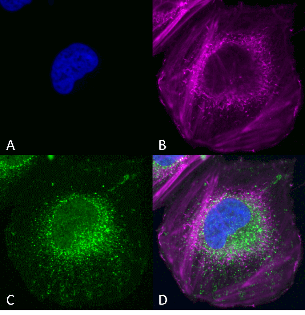
Immunocytochemistry/Immunofluorescence analysis using Rabbit Anti-Beclin 1 Polyclonal Antibody (SPC-601). Tissue: SK-N-BE. Species: Human. Primary Antibody: Rabbit Anti-Beclin 1 Polyclonal Antibody (SPC-601) at 1:200 for Overnight. Secondary Antibody: Anti-Rabbit: AlexaFluor 555 at 1:1000. Counterstain: Hoechst, Phalloidin AlexaFluor 647 at 1:1000. Localization: Mitochondria, endosomes/exosomes . A) Hoechst Nuclear Stain B) Phalloidin AlexaFluor 647 C) Anti-Beclin 1 D) Merge. Courtesy of: Cellstate.
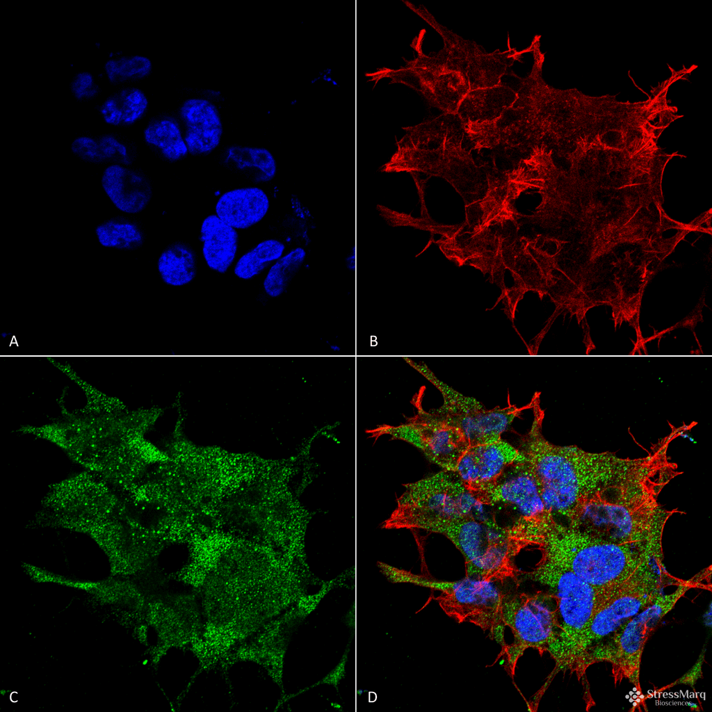
Immunocytochemistry/Immunofluorescence analysis using Rabbit Anti-Beclin 1 Polyclonal Antibody (SPC-601). Tissue: Neuroblastoma cell line (SK-N-BE). Species: Human. Fixation: 4% Formaldehyde for 15 min at RT. Primary Antibody: Rabbit Anti-Beclin 1 Polyclonal Antibody (SPC-601) at 1:100 for 60 min at RT. Secondary Antibody: Goat Anti-Rabbit ATTO 488 at 1:100 for 60 min at RT. Counterstain: Phalloidin Texas Red F-Actin stain; DAPI (blue) nuclear stain at 1:1000, 1:5000 for 60min RT, 5min RT. Localization: Golgi apparatus, Dendrites and Cell bodies of cerebellar Purkinje cells. Magnification: 60X. (A) DAPI (blue) nuclear stain (B) Phalloidin Texas Red F-Actin stain (C) Beclin 1 Antibody (D) Composite.
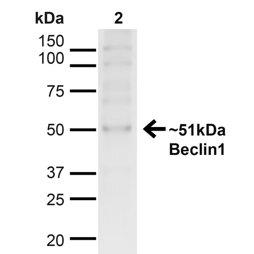
Western blot analysis of Human Cervical cancer cell line (HeLa) lysate showing detection of ~51kDa Beclin 1 protein using Rabbit Anti-Beclin 1 Polyclonal Antibody (SPC-601). Lane 1: MW Ladder. Lane 2: Human HeLa (20 µg). Load: 20 µg. Block: 5% milk + TBST for 1 hour at RT. Primary Antibody: Rabbit Anti-Beclin 1 Polyclonal Antibody (SPC-601) at 1:1000 for 1 hour at RT. Secondary Antibody: Goat Anti-Rabbit: HRP at 1:2000 for 1 hour at RT. Color Development: TMB solution for 12 min at RT. Predicted/Observed Size: ~51kDa.
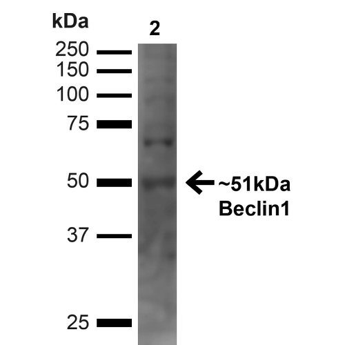
Western blot analysis of Human Embryonic kidney epithelial cell line (HEK293T) lysate showing detection of ~51kDa Beclin 1 protein using Rabbit Anti-Beclin 1 Polyclonal Antibody (SPC-601). Lane 1: MW Ladder. Lane 2: Human 293T (20 µg). Load: 20 µg. Block: 5% milk + TBST for 1 hour at RT. Primary Antibody: Rabbit Anti-Beclin 1 Polyclonal Antibody (SPC-601) at 1:1000 for 1 hour at RT. Secondary Antibody: Goat Anti-Rabbit: HRP at 1:2000 for 1 hour at RT. Color Development: TMB solution for 12 min at RT. Predicted/Observed Size: ~51kDa.
 Powered by Bioz
Reviews
There are no reviews yet.