Discovery through Partnership | Excellence through Quality


Properties
| Storage Buffer | PBS pH7.4, 50% glycerol, 0.09% sodium azide *Storage buffer may change when conjugated |
| Storage Temperature | -20ºC, Conjugated antibodies should be stored according to the product label |
| Shipping Temperature | Blue Ice or 4ºC |
| Purification | Peptide Affinity Purified |
| Clonality | Polyclonal |
| Specificity | Detects ~94kDa. |
| Cite This Product | GRP94 Antibody (StressMarq Biosciences | Victoria, BC CANADA, Catalog# SPC-101, RRID: AB_2703142) |
| Certificate of Analysis | 1 µg/ml of SPC-101 was sufficient for detection of Grp94 in 20 µg of Hela lysate by colorimetric immunoblot analysis using goat anti-rabbit IgG:HRP as the secondary antibody. |
Biological Description
| Alternative Names | HSP90B1, GP96, TRA1, ECGP, 94 kDa glucose regulated protein, 94 kDa glucose-regulated protein, Endoplasmin, Endothelial cell (HBMEC) glycoprotein, ENPL_HUMAN, Glucose regulated protein 94kDa, gp96, gp96 homolog, GRP 94, GRP-94, Heat shock protein 90 kDa beta member 1, heat shock protein 90kDa beta (Grp94), member 1, Heat shock protein, 90 kDa, beta, 1, Stress inducible tumor rejection antigen GP96, tumor rejection antigen (gp96) 1, Tumor rejection antigen 1, Tumor rejection antigen gp96, Tumor rejection antigen-1 (gp96) |
| Research Areas | Cancer, Cancer Metabolism, Cell Signaling, ER Proteins, Heat Shock, Hypoxia, Metabolism, Metabolism processes, Organelle Markers, Protein Trafficking, Response to Hypoxia, Tags and Cell Markers |
| Cellular Localization | Endoplasmic Reticulum, Endoplasmic reticulum lumen, Melanosome |
| Accession Number | NP_035761.1 |
| Gene ID | 22027 |
| Swiss Prot | P08113 |
| Scientific Background |
GRP94 (also known as gp96) is a constitutively expressed ER-resident chaperone and a member of the HSP90 family. It is upregulated in response to cellular stressors such as heat shock, oxidative damage, and glucose deprivation—conditions frequently observed in neurodegenerative diseases. Functionally, GRP94 supports protein translocation, folding, and quality control within the ER. It also plays a key role in immune regulation by facilitating antigen presentation through the MHC class I pathway, linking ER stress to immune responses. In the nervous system, GRP94 contributes to proteostasis and immune surveillance, both of which are disrupted in diseases like Alzheimer’s and Parkinson’s. Structurally, GRP94 forms homodimers and contains a KDEL sequence that ensures ER retention. Despite its homology with cytosolic HSP90, GRP94 exhibits distinct ATP-binding and hydrolysis characteristics, making it a unique therapeutic target. Its dual role in stress adaptation and immune signaling positions GRP94 as a valuable biomarker and potential intervention point in neurodegenerative disease research. |
| References |
1. Rudolph R.W., and Bedows E. (1997) J Biol Chem 272: 3125-3128. 2. Srivastava P.K., et al. (1994) Immunogenetics. 39(2):93-98. 3. Mazzarella R.A., and Green M. (1987) J Biol Chem 262: 8875-8883. 4. Kang, H.S. and Welch W.J. (1991) J Biol Chem 266(9): 5643-5649. 5. Soldano K.L., et al. (2003) J Biol Chem 278(48): 48330-48338. 6. Chu F., et al. (2006) Protein Sci 15(6): 1260-1269. 7. Peter F., et al., (1992) J Biol Chem 267: 10631-10637. 8. Allen S. et al. (2000) Blood 96(2): 560-568. 9. Sato K et al. (2001) Blood 98(6): 1852-1857. 10. Yun S.-W. et al (2000) Brain Research Bulletin.52(5): 371-378. 11. Choukhi A., et al. (1998) J. Virol. 72: 3851-3858. 12. Hoshino T., et al. (1998) Blood 91(11): 4379-4386. 13. Riera M. et al. (1999) Mol. Cell Biochem. 191: 97-104. 14. Gusarova V., et al. (2001) J. Biol. Chem. 276(27): 24891-24900. |
Product Images
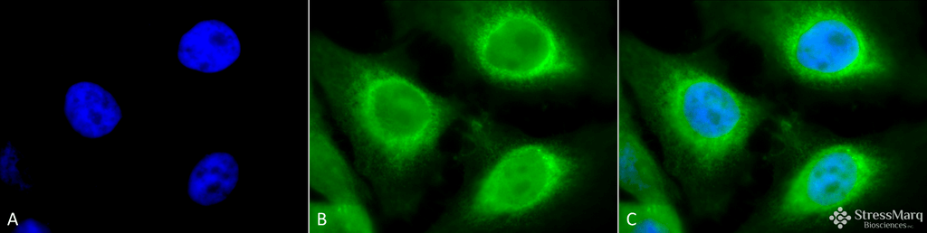
Immunocytochemistry/Immunofluorescence analysis using Rabbit Anti-GRP94 Polyclonal Antibody (SPC-101). Tissue: Heat Shocked Cervical cancer cell line (HeLa). Species: Human. Fixation: 2% Formaldehyde for 20 min at RT. Primary Antibody: Rabbit Anti-GRP94 Polyclonal Antibody (SPC-101) at 1:120 for 12 hours at 4°C. Secondary Antibody: FITC Goat Anti-Rabbit (green) at 1:200 for 2 hours at RT. Counterstain: DAPI (blue) nuclear stain at 1:40000 for 2 hours at RT. Localization: Endoplasmic reticulum lumen. Melanosome. Magnification: 100x. (A) DAPI (blue) nuclear stain. (B) Anti-GRP94 Antibody. (C) Composite. Heat Shocked at 42°C for 1h.
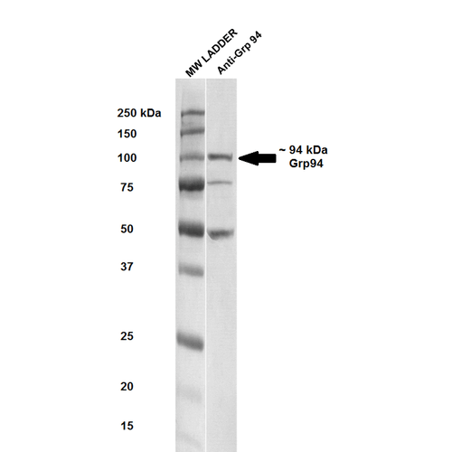
Western blot analysis of Rat brain cell lysates showing detection of ~ 94-100 kDa GRP94 protein using Rabbit Anti-GRP94 Polyclonal Antibody (SPC-101). Lane 1: MW ladder. Lane 2: Anti-GRP94 (1:250). Load: 20 µg. Block: 5% milk + TBST for 1 hour at RT. Primary Antibody: Rabbit Anti-GRP94 Polyclonal Antibody (SPC-101) at 1:250 for 1 hour at RT. Secondary Antibody: Goat Anti-Rabbit HRP antibody at 1:50-1:100 for 1 hour at RT. Color Development: TMB solution for 5 min at RT. Predicted/Observed Size: ~ 94-100 kDa. Other Band(s): ~50, ~75 kDa.
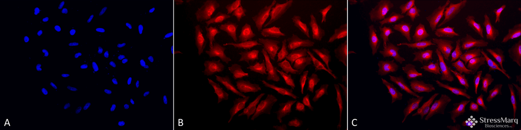
Immunocytochemistry/Immunofluorescence analysis using Rabbit Anti-GRP94 Polyclonal Antibody (SPC-101). Tissue: Heat Shocked Cervical cancer cell line (HeLa). Species: Human. Fixation: 2% Formaldehyde for 20 min at RT. Primary Antibody: Rabbit Anti-GRP94 Polyclonal Antibody (SPC-101) at 1:120 for 12 hours at 4°C. Secondary Antibody: APC Goat Anti-Rabbit (red) at 1:200 for 2 hours at RT. Counterstain: DAPI (blue) nuclear stain at 1:40000 for 2 hours at RT. Localization: Endoplasmic reticulum lumen. Melanosome. Magnification: 20x. (A) DAPI (blue) nuclear stain. (B) Anti-GRP94 Antibody. (C) Composite. Heat Shocked at 42°C for 1h.
 Powered by Bioz
Sepideh :
great for western blot on mouse neuron derived cell lines