Discovery through Partnership | Excellence through Quality


Properties
| Storage Buffer | Rabbit Antiserum *Storage buffer may change when conjugated |
| Storage Temperature | -20ºC, Conjugated antibodies should be stored according to the product label |
| Shipping Temperature | Blue Ice or 4ºC |
| Purification | Rabbit antiserum |
| Clonality | Polyclonal |
| Specificity | Detects ~40kDa. |
| Cite This Product | HSP40 Antibody (StressMarq Biosciences | Victoria, BC CANADA, Catalog# SPC-100, RRID: AB_2261861) |
| Certificate of Analysis | 0.5 µg/ml of SPC-100 was sufficient for detection of HSP40 in 20 µg of heat shocked HeLa cell lysate by colorimetric immunoblot analysis using Goat anti-rabbit IgG:HRP as the secondary antibody. |
Biological Description
| Alternative Names | DNAJB1, DNAJ1, HDJ1, HSPF1, HSP40, DnaJ homolog subfamily B member 1, DnaJ protein homolog 1, Heat shock 40 kDa protein 1, Human DnaJ protein 1, hDj-1, DnaJ (HSP40) homolog subfamily B member 1 |
| Research Areas | Cancer, Cell Signaling, Chaperone Proteins, Heat Shock, Protein Trafficking |
| Cellular Localization | Cytoplasm, Nucleus |
| Accession Number | NP_006136.1 |
| Gene ID | 3337 |
| Swiss Prot | P25685 |
| Scientific Background |
HSP40 proteins, also known as DnaJ homologs, are evolutionarily conserved molecular co-chaperones essential for maintaining protein homeostasis. Characterized by a conserved J-domain within their N-terminal region, these proteins bind to HSP70s and stimulate their ATPase activity—an essential step for HSP70-mediated protein folding, unfolding, translocation, and degradation. By stabilizing the interaction between HSP70 and its substrate proteins, HSP40s effectively determine the functional activity of the HSP70 chaperone system. This interaction is central to the cellular response to proteotoxic stress, particularly in neurons, where the accumulation of misfolded proteins is a hallmark of neurodegenerative diseases such as Alzheimer’s, Parkinson’s, and Huntington’s disease. HSP40 (HDJ1), a 40 kDa mammalian protein, is homologous to bacterial DnaJ and yeast proteins such as YDJ1 and SIS1. It is inducible by stress and translocates from the cytoplasm to the nucleus and nucleoli, mirroring the behavior of HSP70 family members. This dynamic localization suggests a role in nuclear protein quality control and stress signaling. Through its regulation of HSP70 and participation in multi-chaperone complexes, HSP40 is a critical modulator of neuronal proteostasis. Its dysfunction or dysregulation may contribute to the pathogenesis of neurodegenerative disorders, making it a promising target for therapeutic intervention in neuroscience. |
| References |
1. Melville, M. W. et al. (1997) PNAS USA, 94: 97-102. 2. Hattori, H., Liu, Y-C., Tohnai, I., Ueda, M., Kaneda, T., Kobayashi, T., Tanabe, K., and Ohtsuka, K. (1992) Cell Structure and Function 17: 77-86. 3. Ohtsuka, K. Masuda, A., Nakai, A., and Nagata, K. (1990) Biochem. Biophys. Res. Commun. 166: 642-647. 4. Bardwell, J.C.A., Tilly, K., Craig, E., King, J., Zylicz, M. and Georgopoulos, C. (1986) J. Biol. Chem. 261: 1782-1785. 5. Ohku, M., Tamura, F., Nishimura, S., and Uchida, H. (1986) J. Biol. Chem. 261: 1778-1781. 6. Ohtsuka, K. (1993) Biochem. Biophys. Res. Commun. 197: 235-240. |
Product Images
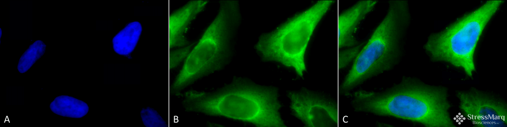
Immunocytochemistry/Immunofluorescence analysis using Rabbit Anti-Hsp40 Polyclonal Antibody (SPC-100). Tissue: Heat Shocked Cervical cancer cell line (HeLa). Species: Human. Fixation: 2% Formaldehyde for 20 min at RT. Primary Antibody: Rabbit Anti-Hsp40 Polyclonal Antibody (SPC-100) at 1:100 for 12 hours at 4°C. Secondary Antibody: FITC Goat Anti-Rabbit (green) at 1:200 for 2 hours at RT. Counterstain: DAPI (blue) nuclear stain at 1:40000 for 2 hours at RT. Localization: Cytoplasm. Magnification: 100x. (A) DAPI (blue) nuclear stain. (B) Anti-Hsp40 Antibody. (C) Composite. Heat Shocked at 42°C for 1h.
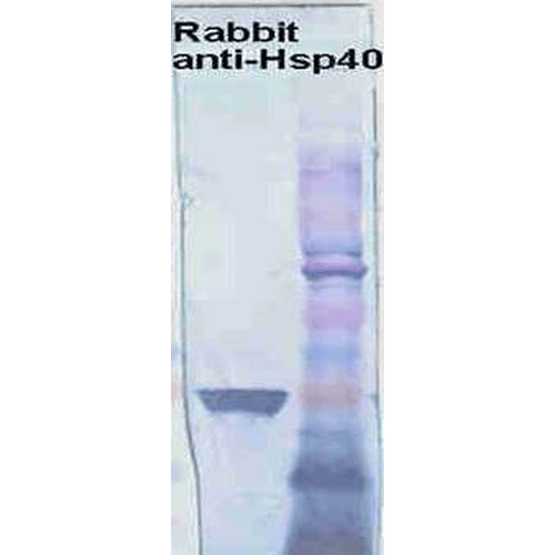
Western blot analysis of Human Cervical cancer cell line (HeLa) lysate showing detection of HSP40 protein using Rabbit Anti-HSP40 Polyclonal Antibody (SPC-100). Primary Antibody: Rabbit Anti-HSP40 Polyclonal Antibody (SPC-100) at 1:1000.
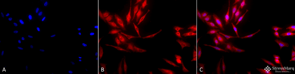
Immunocytochemistry/Immunofluorescence analysis using Rabbit Anti-Hsp40 Polyclonal Antibody (SPC-100). Tissue: Heat Shocked Cervical cancer cell line (HeLa). Species: Human. Fixation: 2% Formaldehyde for 20 min at RT. Primary Antibody: Rabbit Anti-Hsp40 Polyclonal Antibody (SPC-100) at 1:100 for 12 hours at 4°C. Secondary Antibody: APC Goat Anti-Rabbit (red) at 1:200 for 2 hours at RT. Counterstain: DAPI (blue) nuclear stain at 1:40000 for 2 hours at RT. Localization: Cytoplasm. Magnification: 20x. (A) DAPI (blue) nuclear stain. (B) Anti-Hsp40 Antibody. (C) Composite. Heat Shocked at 42°C for 1h.
 Powered by Bioz
StressMarq Biosciences :
Based on validation through cited publications.