Discovery through Partnership | Excellence through Quality


Properties
| Storage Buffer | PBS pH7.4, 50% glycerol, 0.09% sodium azide *Storage buffer may change when conjugated |
| Storage Temperature | -20ºC, Conjugated antibodies should be stored according to the product label |
| Shipping Temperature | Blue Ice or 4ºC |
| Purification | Peptide Affinity Purified |
| Clonality | Polyclonal |
| Specificity | Detects a ~70kDa. May cross-react with HSC70 at lower dilutions. |
| Cite This Product | HSP70 Antibody (StressMarq Biosciences | Victoria, BC CANADA, Catalog# SPC-103, RRID: AB_2119380) |
| Certificate of Analysis | A 1:1000 dilution of SPC-103 was sufficient for detection of HSP70 in 20 µg of HeLa cell lysate by ECL immunoblot analysis. |
Biological Description
| Alternative Names | HSPA1A, HSPA1B, HSPA1, HSP70, HSP70-1, HSP70.1, HSP70-2, HSP72, HSP73, HSX70, Heat shock 70 kDa protein 1A, Heat shock 70 kDa protein 1B |
| Research Areas | Cancer, Cell Signaling, Chaperone Proteins, Heat Shock, Protein Trafficking, Tumor Biomarkers |
| Cellular Localization | Cytoplasm |
| Accession Number | NP_005336.3 |
| Gene ID | 3303 |
| Swiss Prot | P0DMV8/P0DMV9 |
| Scientific Background |
HSP70 proteins are a highly conserved family of 70-kDa molecular chaperones encoded by a multigene family in most eukaryotes and prokaryotes. Found in nearly all cellular compartments—including the cytosol, nucleus, mitochondria, endoplasmic reticulum, and chloroplasts—HSP70s are constitutively expressed and strongly upregulated in response to cellular stress. These chaperones play a vital role in protein homeostasis by binding to nascent polypeptides and partially folded or misfolded proteins, preventing aggregation and facilitating proper folding. HSP70s exhibit high-affinity ATP binding and weak ATPase activity, which is stimulated upon interaction with unfolded substrates. ATP hydrolysis triggers conformational changes that regulate substrate binding and release, enabling dynamic cycles of protein folding and refolding. Structurally, the N-terminal domain of HSP70 is responsible for ATP binding, while the C-terminal domain mediates substrate interaction. This modular design allows HSP70s to coordinate with co-chaperones such as HSP40, HIP, HOP, and BAG-1, integrating folding with degradation and transport pathways. In neurodegenerative disease research, HSP70 is of particular interest due to its ability to counteract protein misfolding and aggregation—hallmarks of disorders like Alzheimer’s, Parkinson’s, and Huntington’s disease. By stabilizing toxic intermediates and promoting their clearance, HSP70 enhances neuronal survival and resilience under proteotoxic stress. |
| References |
1. Welch W.J. and Suhan J.P. (1986) J.Cell Biol. 103: 2035-2050. 2. Boorstein W. R., Ziegelhoffer T. & Craig E. A. (1993) J. Mol. Evol. 38(1): 1-17. 3. Rothman J. (1989) Cell 59: 591 -601. 4. DeLuca-Flaherty et al. (1990) Cell 62: 875-887. 5. Bork P., Sander C. & Valencia A. (1992) Proc. Nut1 Acad. Sci. USA 89: 7290-7294. 6. Fink A.L. (1999) Physiol. Rev. 79: 425-449. 7. Hung T.H., et al. (2001) Am J Pathol. 159: 1031-1043. 8. Locke M. (2000) Cell Stress & Chaperones 5: 45-51. 9. Ianaro A., et al. (2001) FEBS Lett. 508: 61-66. 10.Trentin G.A. et al. (2001) J Biol Chem. 276: 13087-13095. |
Product Images
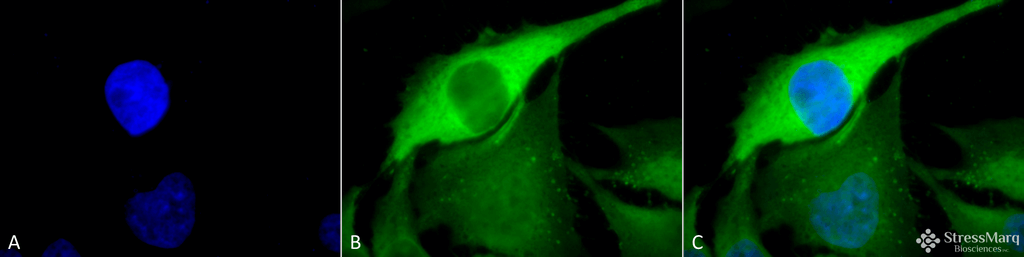
Immunocytochemistry/Immunofluorescence analysis using Rabbit Anti-Hsp70 Polyclonal Antibody (SPC-103). Tissue: Heat Shocked Cervical cancer cell line (HeLa). Species: Human. Fixation: 2% Formaldehyde for 20 min at RT. Primary Antibody: Rabbit Anti-Hsp70 Polyclonal Antibody (SPC-103) at 1:100 for 12 hours at 4°C. Secondary Antibody: FITC Goat Anti-Rabbit (green) at 1:200 for 2 hours at RT. Counterstain: DAPI (blue) nuclear stain at 1:40000 for 2 hours at RT. Localization: Cytoplasm. Magnification: 100x. (A) DAPI (blue) nuclear stain. (B) Anti-Hsp70 Antibody. (C) Composite. Heat Shocked at 42°C for 1h.
Immunohistochemistry analysis using Rabbit Anti-HSP70 Polyclonal Antibody (SPC-103). Tissue: colon carcinoma. Species: Human. Fixation: Formalin. Primary Antibody: Rabbit Anti-HSP70 Polyclonal Antibody (SPC-103) at 1:50000 for 12 hours at 4°C. Secondary Antibody: Biotin Goat Anti-Rabbit at 1:2000 for 1 hour at RT. Counterstain: Methyl Green at 200uL for 2 min at RT.
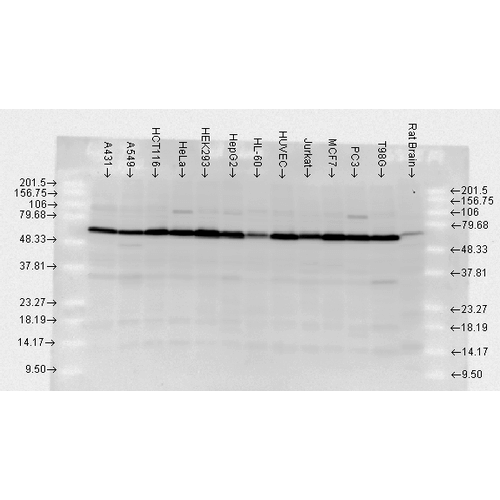
Western blot analysis of Human, Rat brain cell lysates showing detection of HSP70 protein using Rabbit Anti-HSP70 Polyclonal Antibody (SPC-103). Load: 2 µg. Block: 1.5% BSA for 30 minutes at RT. Primary Antibody: Rabbit Anti-HSP70 Polyclonal Antibody (SPC-103) at 1:10000 for 2 hours at RT. Secondary Antibody: Donkey Anti-Rabbit IgG: HRP for 1 hour at RT.
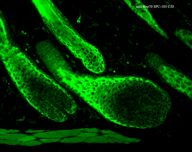
Immunohistochemistry analysis using Rabbit Anti-HSP70 Polyclonal Antibody (SPC-103). Tissue: backskin. Species: Mouse. Fixation: Bouin’s Fixative Solution. Primary Antibody: Rabbit Anti-HSP70 Polyclonal Antibody (SPC-103) at 1:100 for 1 hour at RT. Secondary Antibody: FITC Goat Anti-Rabbit (green) at 1:50 for 1 hour at RT. Localization: Cytoplasm.
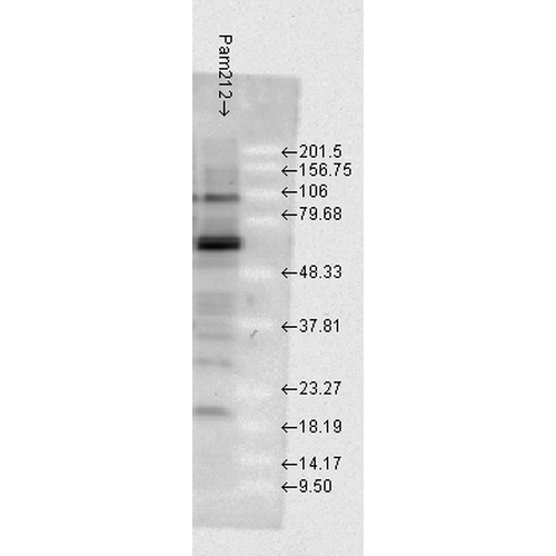
Western blot analysis of Mouse Pam212 cells showing detection of HSP70 protein using Rabbit Anti-HSP70 Polyclonal Antibody (SPC-103). Load: 15 µgprotein. Block: 1.5% BSA for 30 minutes at RT. Primary Antibody: Rabbit Anti-HSP70 Polyclonal Antibody (SPC-103) at 1:1000 for 2 hours at RT. Secondary Antibody: Donkey Anti-Rabbit IgG: HRP for 1 hour at RT.
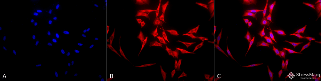
Immunocytochemistry/Immunofluorescence analysis using Rabbit Anti-Hsp70 Polyclonal Antibody (SPC-103). Tissue: Heat Shocked Cervical cancer cell line (HeLa). Species: Human. Fixation: 2% Formaldehyde for 20 min at RT. Primary Antibody: Rabbit Anti-Hsp70 Polyclonal Antibody (SPC-103) at 1:100 for 12 hours at 4°C. Secondary Antibody: APC Goat Anti-Rabbit (red) at 1:200 for 2 hours at RT. Counterstain: DAPI (blue) nuclear stain at 1:40000 for 2 hours at RT. Localization: Cytoplasm. Magnification: 20x. (A) DAPI (blue) nuclear stain. (B) Anti-Hsp70 Antibody. (C) Composite. Heat Shocked at 42°C for 1h.
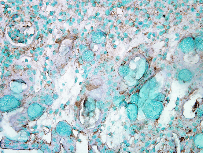
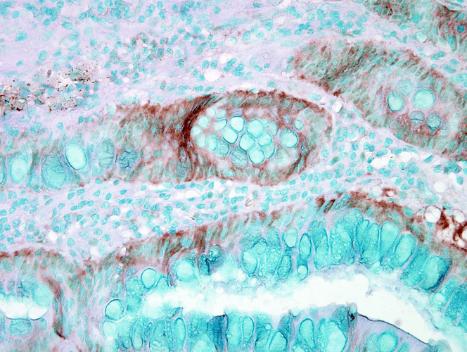
Immunohistochemistry analysis using Rabbit Anti-HSP70 Polyclonal Antibody (SPC-103). Tissue: Inflamed colon. Species: Mouse. Fixation: Formalin. Primary Antibody: Rabbit Anti-HSP70 Polyclonal Antibody (SPC-103) at 1:1000 for 12 hours at 4°C. Secondary Antibody: Biotin Goat Anti-Rabbit at 1:2000 for 1 hour at RT. Counterstain: Methyl Green at 200uL for 2 min at RT.
 Powered by Bioz
StressMarq Biosciences :
Based on validation through cited publications.