Discovery through Partnership | Excellence through Quality


Properties
| Storage Buffer | PBS pH7.4, 50% glycerol, 0.09% sodium azide *Storage buffer may change when conjugated |
| Storage Temperature | -20ºC, Conjugated antibodies should be stored according to the product label |
| Shipping Temperature | Blue Ice or 4ºC |
| Purification | Peptide Affinity Purified |
| Clonality | Polyclonal |
| Specificity | Detects ~55kDa. Other bands present may be the result of oligomerization, self-aggregation and/or cleavage of the TNF-R1 extracellular domain. |
| Cite This Product | TNF-R1 Antibody (StressMarq Biosciences | Victoria, BC CANADA, Catalog# SPC-170, RRID: AB_2703803) |
| Certificate of Analysis | 1 µg/ml of SPC-170 was sufficient for detection of TNFR1 in 20 µg of Hela lysate by colorimetric immunoblot analysis using Goat anti-rabbit IgG:HRP as the secondary antibody. |
Biological Description
| Alternative Names | Tumor necrosis factor receptor 1, TNFR-1, TNFRSF1A, TNFAR, TNFR1 |
| Research Areas | Apoptosis, Atherosclerosis, Cancer, Cardiovascular System, Cell Signaling |
| Cellular Localization | Cell membrane, Golgi apparatus, Golgi Apparatus Membrane |
| Gene ID | 7132 |
| Swiss Prot | P19438 |
| Scientific Background |
Tumor necrosis factor receptor 1 (TNF-R1), also known as CD120a or p55/p60, is a high-affinity receptor for tumor necrosis factor-alpha (TNF-α) and a key member of the TNF/TNFR superfamily. Structurally, TNF-R1 features an extracellular domain with multiple cysteine-rich repeats and a cytoplasmic death domain that initiates downstream signaling cascades. TNF-R1 is widely expressed in the central nervous system, including neurons, astrocytes, and microglia. Upon binding TNF-α, TNF-R1 activates multiple intracellular pathways, including NF-κB-mediated inflammation and caspase-dependent apoptosis. This dual functionality allows TNF-R1 to regulate both cell survival and programmed cell death, depending on the cellular context and signaling environment. In neurodegenerative disease research, TNF-R1 has emerged as a critical mediator of chronic neuroinflammation and neuronal loss. Aberrant TNF-R1 signaling is implicated in the pathogenesis of Alzheimer’s disease, Parkinson’s disease, multiple sclerosis, and amyotrophic lateral sclerosis (ALS). Sustained activation of TNF-R1 can exacerbate oxidative stress, synaptic dysfunction, and glial reactivity—hallmarks of progressive neurodegeneration. Given its central role in immune signaling and apoptosis, TNF-R1 is a promising therapeutic target for modulating neuroinflammatory responses and protecting neuronal integrity in neurodegenerative disorders. |
| References |
1. Kontermann R.E., et al. (2008) J Immunother. 31(3):225-34. 2. Hehlgans T. and Pfeffer K. (2005) Immunology. 115(1):1-20. 3. Al-Lamki S., et al. (2005) The Faseb Journal. 19:1638-1645. |
Product Images
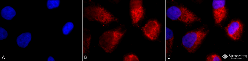
Immunocytochemistry/Immunofluorescence analysis using Rabbit Anti-TNF-R1 Polyclonal Antibody (SPC-170). Tissue: Cervical cancer cell line (HeLa). Species: Human. Fixation: 2% Formaldehyde for 20 min at RT. Primary Antibody: Rabbit Anti-TNF-R1 Polyclonal Antibody (SPC-170) at 1:100 for 12 hours at 4°C. Secondary Antibody: APC Goat Anti-Rabbit (red) at 1:200 for 2 hours at RT. Counterstain: DAPI (blue) nuclear stain at 1:40000 for 2 hours at RT. Localization: Golgi apparatus membrane. Magnification: 100x. (A) DAPI (blue) nuclear stain. (B) Anti-TNF-R1 Antibody. (C) Composite.
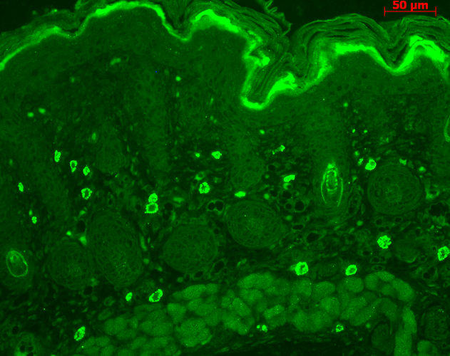
Immunohistochemistry analysis using Rabbit Anti-TNF-R1 Polyclonal Antibody (SPC-170). Tissue: backskin. Species: Mouse. Fixation: Bouin’s Fixative Solution. Primary Antibody: Rabbit Anti-TNF-R1 Polyclonal Antibody (SPC-170) at 1:100 for 1 hour at RT. Secondary Antibody: FITC Goat Anti-Rabbit (green) at 1:50 for 1 hour at RT. Localization: dermis.
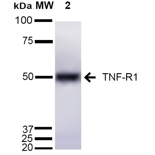
Western blot analysis of Mouse Liver cell lysates showing detection of ~55 kDa TNF-R1 protein using Rabbit Anti-TNF-R1 Polyclonal Antibody (SPC-170). Lane 1: Molecular Weight Ladder (MW). Lane 2: Mouse Liver cell lysates. Load: 15 µg. Block: 5% Skim Milk in 1X TBST. Primary Antibody: Rabbit Anti-TNF-R1 Polyclonal Antibody (SPC-170) at 1:1000 for 2 hours at RT. Secondary Antibody: Goat Anti-Rabbit IgG: HRP at 1:2000 for 60 min at RT. Color Development: ECL solution for 5 min at RT. Predicted/Observed Size: ~55 kDa.
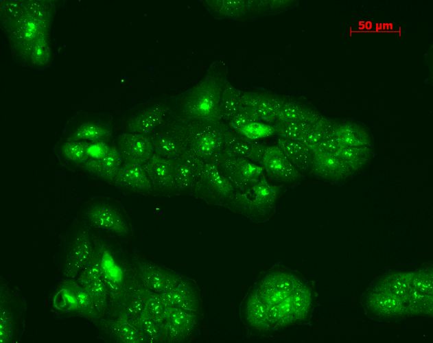
Immunocytochemistry/Immunofluorescence analysis using Rabbit Anti-TNF-R1 Polyclonal Antibody (SPC-170). Tissue: HaCaT cells. Species: Human. Fixation: Cold 100% methanol at -20C for 10 minutes. Primary Antibody: Rabbit Anti-TNF-R1 Polyclonal Antibody (SPC-170) at 1:100 for 12 hours at 4°C. Secondary Antibody: FITC Goat Anti-Rabbit at 1:50 for 1-2 hours at RT in dark. Localization: Punctate nuclear staining, dotty staining in cytoplasm.
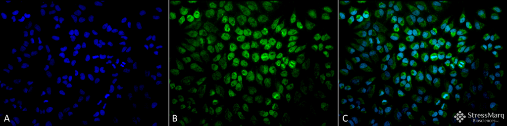
Immunocytochemistry/Immunofluorescence analysis using Rabbit Anti-TNF-R1 Polyclonal Antibody (SPC-170). Tissue: Cervical cancer cell line (HeLa). Species: Human. Fixation: 2% Formaldehyde for 20 min at RT. Primary Antibody: Rabbit Anti-TNF-R1 Polyclonal Antibody (SPC-170) at 1:100 for 12 hours at 4°C. Secondary Antibody: FITC Goat Anti-Rabbit (green) at 1:200 for 2 hours at RT. Counterstain: DAPI (blue) nuclear stain at 1:40000 for 2 hours at RT. Localization: Golgi apparatus membrane. Magnification: 20x. (A) DAPI (blue) nuclear stain. (B) Anti-TNF-R1 Antibody. (C) Composite.
 Powered by Bioz
Reviews
There are no reviews yet.