Discovery through Partnership | Excellence through Quality


Properties
| Storage Buffer | PBS, 50% glycerol, 0.09% sodium azide *Storage buffer may change when conjugated |
| Storage Temperature | -20ºC, Conjugated antibodies should be stored according to the product label |
| Shipping Temperature | Blue Ice or 4ºC |
| Purification | Peptide Affinity Purified |
| Clonality | Polyclonal |
| Specificity | Detects ~100 kDa. |
| Cite This Product | ULK1 Antibody (StressMarq Biosciences | Victoria, BC CANADA, Catalog# SPC-640, RRID: AB_2705267) |
| Certificate of Analysis | A 1:1000 dilution of SPC-640 was sufficient for detection of ULK1 in 15 µg of Human HeLa Cell Lysates by ECL immunoblot analysis using goat anti-rabbit IgG:HRP as the secondary antibody. |
Biological Description
| Alternative Names | Unc-51 like kinase 1 (C. elegans), Autophagy related protein 1 homolog, Autophagy-related protein 1 homolog, FLJ38455, Serine/threonine-protein kinase ULK1, Unc51.1, ATG1A, UNC51, C. elegans, homolog of, EC:2.7.11.1, Serine/threonine protein kinase Unc51.1, FLJ46475, ATG 1, hATG1, ATG1, Unc-51-like kinase 1, Serine/threonine protein kinase ULK1, ULK1, Unc 51 like kinase 1, KIAA0722, UNC 51, ULK 1, ULK1_HUMAN, UNC51, Unc 51 (C. elegans) like kinase 1, ATG1 autophagy related 1 homolog |
| Research Areas | Cardiovascular System, Cell Signaling, Heart, Metabolism, Mitochondrial Metabolism, Neuroscience, Protein Phosphorylation, Serine / Threonine Kinases |
| Cellular Localization | Cytoplasm, Cytosol, Preautophagosomal structure |
| Accession Number | NP_003556.1 |
| Gene ID | 8408 |
| Swiss Prot | O75385 |
| Scientific Background |
UNC-51-like kinase 1 (ULK1) is a serine/threonine kinase broadly expressed in mammalian tissues, with a critical role in neuronal development and homeostasis. Structurally, ULK1 comprises an N-terminal kinase domain, a central proline/serine-rich region, and a highly conserved C-terminal domain. Functionally, ULK1 is a mammalian homolog of yeast Atg1 and serves as a master regulator of autophagy initiation—a process essential for cellular quality control and survival under stress. In the nervous system, ULK1 has been directly implicated in axonal growth, synaptic remodeling, and neuronal survival. Its ability to interact with multiple autophagy-related (ATG) proteins enables it to orchestrate phosphorylation-dependent signaling cascades that regulate autophagosome formation and intracellular trafficking. Dysregulation of ULK1 activity has been associated with impaired autophagic flux, a hallmark of several neurodegenerative diseases including Alzheimer’s, Parkinson’s, and Huntington’s disease. Given its dual role in autophagy and axonal dynamics, ULK1 represents a promising therapeutic target in neuroscience and neurodegeneration research. Understanding ULK1-mediated signaling pathways offers critical insights into the molecular mechanisms underlying neuronal dysfunction and opens new avenues for the development of autophagy-based interventions in neurodegenerative disorders. |
| References |
1. Okazaki N., et al. (2000) Brain Res Mol Brain Res. 85: 1-12. 2. Young A.R., et al. (2006) J Cell Sci. 119: 3888-900. 3. Kamada Y., et al. (2000) J Cell Biol. 150: 1507-13. 4. Lee S.B, et al. (2007) EMBO Rep. 8: 360-5. 5. Hara T., et al. (2008) J Cell Biol. 181: 497-510. |
Product Images
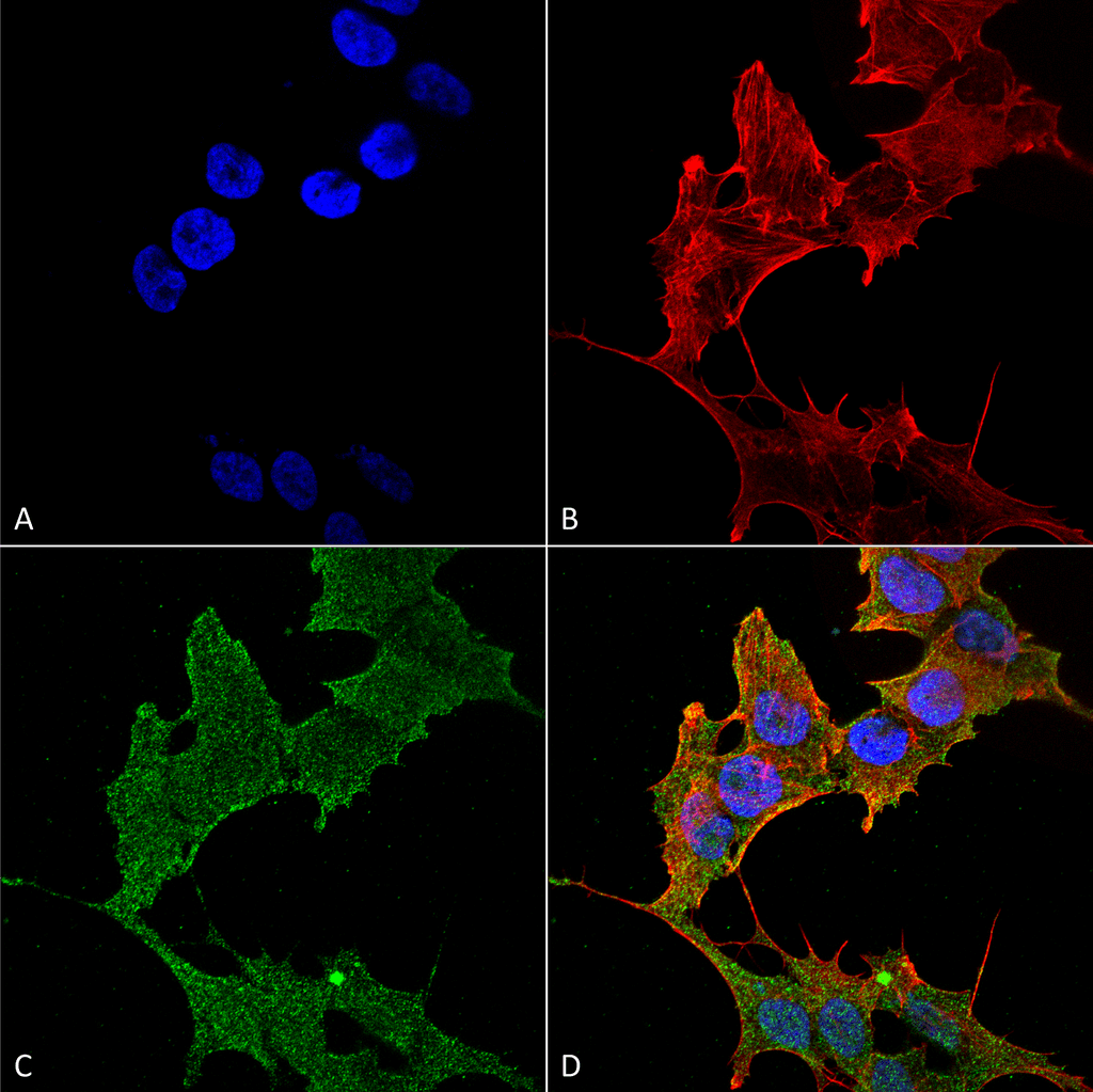
Immunocytochemistry/Immunofluorescence analysis using Rabbit Anti-ULK1 Polyclonal Antibody (SPC-640). Tissue: Neuroblastoma cell line (SK-N-BE). Species: Human. Fixation: 4% Formaldehyde for 15 min at RT. Primary Antibody: Rabbit Anti-ULK1 Polyclonal Antibody (SPC-640) at 1:100 for 60 min at RT. Secondary Antibody: Goat Anti-Rabbit ATTO 488 at 1:200 for 60 min at RT. Counterstain: Phalloidin Texas Red F-Actin stain; DAPI (blue) nuclear stain at 1:1000, 1:5000 for 60 min at RT, 5 min at RT. Localization: Cytoplasm, Preautophagosomal Structure. Magnification: 60X. (A) DAPI (blue) nuclear stain (B) Phalloidin Texas Red F-Actin stain (C) ULK1 Antibody (D) Composite.
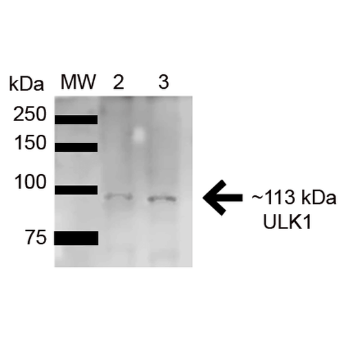
Western blot analysis of Human HeLa and HEK293Trap cell lysates showing detection of 112.6 kDa ULK1 protein using Rabbit Anti-ULK1 Polyclonal Antibody (SPC-640). Lane 1: Molecular Weight Ladder (MW). Lane 2: HeLa (15ug) . Lane 3: Human 293Trap (15ug) . Load: 15 µg. Block: 5% Skim Milk in 1X TBST. Primary Antibody: Rabbit Anti-ULK1 Polyclonal Antibody (SPC-640) at 1:1000 for 1 hour at RT. Secondary Antibody: Goat Anti-Rabbit HRP at 1:2000 for 60 min at RT. Color Development: ECL solution for 6 min in RT. Predicted/Observed Size: 112.6 kDa.
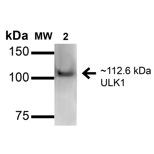
Western blot analysis of Rat Brain cell lysates showing detection of ~112.6kDa ULK1 protein using Rabbit Anti-ULK1 Polyclonal Antibody (SPC-640). Lane 1: Molecular Weight Ladder (MW). Lane 2: Rat Brain cell lysates. Load: 15 µg. Block: 2% GE Healthcare Blocker (RT, 60 minutes). Primary Antibody: Rabbit Anti-ULK1 Polyclonal Antibody (SPC-640) at 1:1000 for 16 hours at 4°C. Secondary Antibody: Goat Anti-Rabbit IgG: HRP at 1/2000 for 60 min at RT. Color Development: ECL solution for 6 min at RT. Predicted/Observed Size: ~112.6kDa.
 Powered by Bioz
Reviews
There are no reviews yet.