Discovery through Partnership | Excellence through Quality
| Product Name | Corticosterone EIA Kit |
| Description |
Colorimetric detection of corticosterone |
| Species Reactivity | Species Independent |
| Platform | Microplate |
| Sample Types | EDTA Plasma, Heparin Plasma, Serum, Tissue Culture Media, Urine |
| Detection Method | Colorimetric Assay |
| Assay Type | Sandwich EIA (Enzyme Immunoassay) |
| Utility | EIA kit used to measure the corticosterone present in samples. |
| Sensitivity | 24.7 pg/mL |
| Assay Range | 78.125 - 10,000 pg/ml |
| Precision | Intra Assay Precision: Four human samples were diluted with Assay Buffer and run in replicates of 24 in an assay. The mean and precision of the calculated Corticosterone concentrations were: Sample 1- 322.5 pg/mL, 2.6% CV Sample 2- 564.3 pg/mL, 2.8% CV Sample 3- 630.3 pg/mL, 3.1% CV Inter Assay Precision: Three human samples were diluted with Assay Buffer and run in replicates of 24 in two assays. The mean and precision of the calculated Corticosterone concentrations were: Sample 1- 732.6 pg/mL, 1.6% CV Sample 2- 402.7 pg/mL, 0.5% CV Sample 3- 594.5 pg/mL, 0.2% CV |
| Number of Samples | 39 samples in duplicate |
| Other Resources | Kit Booklet Lot No. SC332123 , Kit Booklet Lot No. SC989258 , Kit Booklet Lot No. SC188662 , MSDS , Steroid Solid Extraction Protocol |
| Field of Use | Not for use in humans. Not for use in diagnostics or therapeutics. For in vitro research use only. |
Properties
| Storage Temperature | 4ºC and -20ºC | ||||||||||||||||||||||||||||||||||||
| Shipping Temperature | Blue Ice | ||||||||||||||||||||||||||||||||||||
| Product Type | EIA Kits | ||||||||||||||||||||||||||||||||||||
| Assay Overview | The Corticosterone EIA kit is designed to quantitatively measure Corticosterone present in serum, plasma, urine, extracted dried fecal samples, and tissue culture media samples. This kit measures total corticosterone in serum and plasma and in extracted fecal samples. A corticosterone stock solution is provided to generate a standard curve for the assay and all samples should be read off the standard curve. We provide protocols on page 8 to prepare assay standards from 5,000 to 78.125 pg/mL or from 10,000 to 78.125 pg/mL. Please choose the standard range that fits your sample concentrations most appropriately. Standards or diluted samples are pipetted into a clear microtiter plate coated with an antibody to capture sheep antibodies. A corticosterone-peroxidase conjugate is added to the standards and samples in the wells. The binding reaction is initiated by the addition of a polyclonal antibody to corticosterone to each well. After an hour incubation the plate is washed and substrate is added. The substrate reacts with the bound corticosterone-peroxidase conjugate. After a short incubation, the reaction is stopped and the intensity of the generated color is detected in a microtiter plate reader capable of measuring 450nm wavelength. The concentration of the corticosterone in the sample is calculated, after making suitable correction for the dilution of the sample, using software available with most plate readers. | ||||||||||||||||||||||||||||||||||||
| Kit Overview |
|
||||||||||||||||||||||||||||||||||||
| Cite This Product | Corticosterone EIA Kit (StressMarq Biosciences Inc., Victoria BC CANADA, Catalog # SKT-205) |
Biological Description
| Alternative Names | (11β)-11,21-dihydroxypregn-4-ene-3,20-dione |
| Research Areas | Cancer, Cell Signaling, Neuroscience, Oxidative Stress |
| Scientific Background |
Corticosterone (C₂₁H₃₀O₄) is a glucocorticoid hormone secreted by the adrenal cortex in response to ACTH stimulation. It is the primary stress hormone in rodents and a precursor to aldosterone. In neuroscience research, corticosterone is widely used to model chronic stress and its effects on brain function. Elevated corticosterone levels have been shown to impair memory retrieval, alter sleep-wake cycles, and exacerbate neurodegenerative processes. In Parkinson’s disease models, chronic corticosterone exposure increases alpha-synuclein phosphorylation and aggregation, particularly in the hypothalamus, and contributes to dopaminergic neuron loss. These findings highlight corticosterone’s role in stress-induced neurotoxicity and its relevance in studying the pathophysiology of neurodegenerative and psychiatric disorders. |
| References |
1. Hupé, J. M., James, A. C., Payne, B. R., Lomber, S. G., Girard, P., & Bullier, J. (1998). Cortical feedback improves discrimination between figure and background by V1, V2, and V3 neurons. Nature, 394(6695), 784–787. DOI: 10.1038/29555 2. Kitaysky, A. S., Kitaiskaia, E. V., Wingfield, J. C., & Piatt, J. F. (2001). Dietary restrictions cause chronic elevation of corticosterone and enhance stress response in red-legged kittiwake chicks. Journal of Comparative Physiology B, 171(8), 701–709. DOI: 10.1007/s003600100230 3. Thellin, O., Noel, G., Khuana, S., Ogle, C. K., & Horseman, N. D. (2001). Stress hormone secretion and gut signal transducer (STAT) proteins after burn injury in rats. Shock, 16(5), 393–397. DOI: 10.1097/00024382-200116050-00001 4. Kramer, K. M., & Sothern, R. B. (2001). Circadian characteristics of corticosterone secretion in red-backed voles (Clethrionomys gapperi). Chronobiology International, 18(6), 933–945. DOI: 10.1081/CBI-100107542 5. Vazquez-Palacios, G., Retana-Márquez, S., Bonilla-Jaime, H., & Velázquez-Moctezuma, J. (2001). Further definition of the effect of corticosterone on the sleep-wake pattern in the male rat. Pharmacology, Biochemistry, and Behavior, 70(2–3), 305–310. DOI: 10.1016/S0091-3057(01)00629-8 6. Bimpos, N. M., Xie, Y., Gomez-Isla, T., Domenger, D., & Hyman, B. T. (2024). Alpha-synuclein-induced stress sensitivity renders the Parkinson’s disease brain susceptible to neurodegeneration. Acta Neuropathologica Communications, 12, 100. DOI: 10.1186/s40478-024-01797-w. 7. Burtscher, J., Copin, J. C., Rodrigues, J., & Pernet, N. (2019). Chronic corticosterone aggravates behavioral and neuronal symptomatology in a mouse model of alpha-synuclein pathology. Neurobiology of Aging, 83, 11–20. DOI: 10.1016/j.neurobiolaging.2019.08.011 |
Product Images
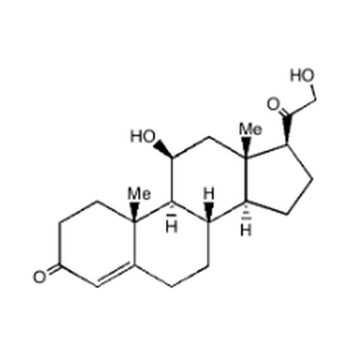
Chemical structure of Corticosterone
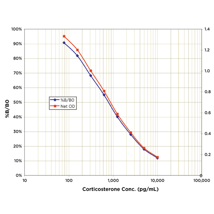
Typical Standard Curve for the Corticosterone EIA Kit (Enzyme Immunoassay) StressXpress® – SKT-205. Assay Type: Sandwich EIA. Detection Method: Colorimetric Assay. Assay Range: 78.125 – 10,000 pg/ml.
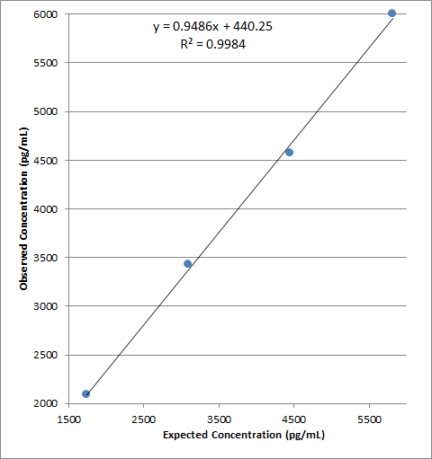
Linearity was determined by taking two serum samples treated with Dissociation Reagent and diluted 1:50 with Assay Buffer, one with a low diluted corticosterone level of 104.6 pg/mL and one with a higher diluted level of 2,890.5 pg/mL, and mixing them in the ratios given below. The measured concentrations were compared to the expected values based on the ratios used.
 Powered by Bioz
StressMarq Biosciences :
Based on validation through cited publications.