Discovery through Partnership | Excellence through Quality
MOLECULAR SIGNATURE®
Dibromo-tyrosine ELISA Kit (Discontinued)
Buy 5 or more to get 15% OFF
 MOLECULAR SIGNATURE® - SKT-140. Assay Type: Competitive ELISA. Detection Method: Colorimetric Assay. Assay Range: 0.078 - 5 µg/ml.")
| Product Name | Dibromo-tyrosine ELISA Kit (Discontinued) |
| Description |
Colorimetric detection of 3,5-dibromotyrosine |
| Species Reactivity | Species Independent |
| Platform | Microplate |
| Sample Types | Plasma, Urine |
| Detection Method | Colorimetric Assay |
| Assay Type | Competitive ELISA (Enzyme-linked Immunosorbent Assay) |
| Utility | ELISA kit used to quantify free and protein-bound 3,5-dibromotyrosine in samples |
| Sensitivity | 0.04 µg/ml |
| Assay Range | 0.078 - 5 µg/mL |
| Precision | Intra-Assay Precision: Three samples of known concentration were assayed thirty times on one plate; the intra-assay coefficient of variation of the Dibromo-tyrosine ELISA has been determined to be < 10%. Inter-Assay Precision: Three samples of known concentration were assayed thirty times in three individual assays; the inter-assay coefficient of variation of the Dibromo-tyrosine ELISA has been determined to be < 10%. |
| Incubation Time | 1 hour |
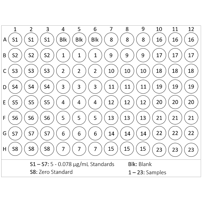
| Number of Samples | 39 samples in duplicate, 23 in triplicate |
| Other Resources | Kit Booklet , MSDS , Calculations Worksheet |
| Field of Use | Not for use in humans. Not for use in diagnostics or therapeutics. For in vitro research use only. |
Properties
| Storage Temperature | 4ºC and -20ºC | ||||||||||||||||||||||||||||||||||||
| Shipping Temperature | Blue Ice | ||||||||||||||||||||||||||||||||||||
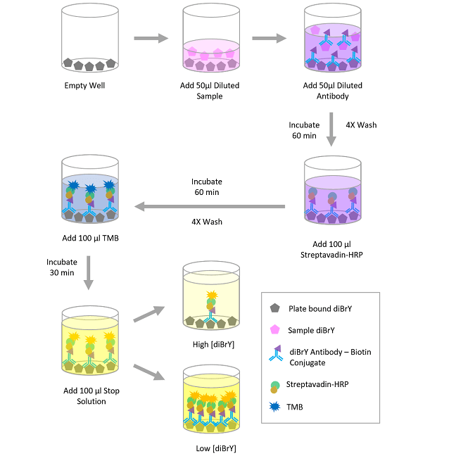
| Assay Overview | 1. Prepare standard and samples in the Sample and Standard Diluent. 2. Add 50 µL of prepared standards and samples in triplicate to appropriate wells. 3. Add 50 µL of the diluted Dibromo-tyrosine-Biotin antibody to the appropriate wells. 4. Cover plate with Plate Cover and incubate at 37 °C for 1 hour. 5. Wash plate 4 times with 1X Wash Buffer. 6. Add 100 μL of Streptavidin-HRP Working Solution to each well. 7. Cover plate with Plate Sealer and incubate at room temperature for 30 minutes. 8. Wash plate 4 times with 1X Wash Buffer. 9. Add 100 µL of TMB Substrate to each well. 10. Develop the plate in the dark at room temperature (20-25°C) for 30 minutes. 11. Stop reaction by adding 100 μL of Stop Solution to each well. 12. Measure absorbance on plate reader at 450 nm. | ||||||||||||||||||||||||||||||||||||
| Kit Overview |
|
||||||||||||||||||||||||||||||||||||
| Cite This Product | Dibromo-tyrosine ELISA Kit (StressMarq Biosciences, Canada, Cat # SKT-140) |
Biological Description
| Alternative Names | 3,5-Dibromo-tyrosine ELISA Kit, 3,5-dibromotyrosine ELISA Kit, Dibromotyrosine ELISA Kit, Dibromo-tyrosine ELISA Kit, DiBrY ELISA Kit, Dibromotyrosine (DiBrY) ELISA Kit, Dibromo-Tyrosine (DiBrY) ELISA Kit, Dibromo-tyrosine [DiBrY] Antibody |
| Research Areas | Cancer, Cell Signaling, Oxidation, Oxidative Stress, Post-translational Modifications, Protein Oxidation |
| Scientific Background |
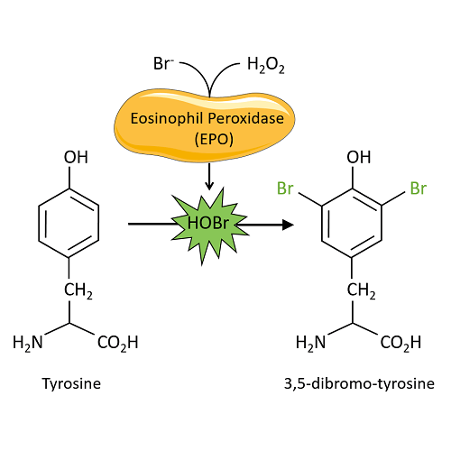
Dibromo-tyrosine is produced by the oxidative bromination of tyrosine residues. This reaction occurs via eosinophil peroxidase (EPO), an enzyme released by activated eosinophils. Upon activation of eosinophils, a respiratory burst occurs releasing elevated levels of O2 and H202. In the oxidation of tyrosine, EPO utilizes H202 to catalyze the peroxidation of physiological levels of bromine found within plasma to generate the brominating reagent hypobromous acid (HOBr)(1-5). Eosinophils play an immunomodulatory role through their recruitment to host sites of parasitic invasion. EPO levels also contribute to diseases such as asthma, cancers and allergic disorders where cellular activation is found to occur at pathological sites (6-10). Brominated products such as 3,5-dibromo-tyrosine serve as biological markers for in vivo eosinophil-mediated tissue damage which allows for understanding the overall roll oxidative stress has on pathways implicated in diseased states within organisms (4). |
| References |
1. MacPherson, J.C., Comhair, S. A. A., Erzurum, S.C., et al. J. Immun. 166, 5763-577 (2001). 2. Mayeno, A. N., Curran, A. J., Roberts, R. L., et al. J. Biol. Chem. 264, 5660-5668 (1989). 3. Babior, B. M. N. Engl. J. Med. 298, 659-668 (1978). 4. Wu W., Chen, Y., d’Avignon, A. et al. Biochem. 38, 3538-3548 (1999). 5. Kambayashi, Y., Ogino, K., Takemoto, K. et al. J. Clin. Biochem. Nutr. 44, 95-103 (2009). 6. Wang J., Slungaard A. Arch. Biochem. Biophys. 445, 256–260 (2006). 7. Kazura, J. W., Fanning, M. M., Blumer, J. L. Mahmoud, A. A. J. Clin. Invest. 67, 93 (1981). 8. Klebanoff, S. J., Locksley, R. M., Jong, E. C., Rosen, H. CIBA Found. Symp. 99: 92 (1983). 9. Gleich, G. J., Ottesen, E. A., Leiferman, K. M., Ackerman, S. J. Int. Arc. Allergy Appl. Immunol. 88: 59 (1989). 10. Wardlaw, A. J., Postgrad. Med. J. 70: 536 (1994). |
Product Images
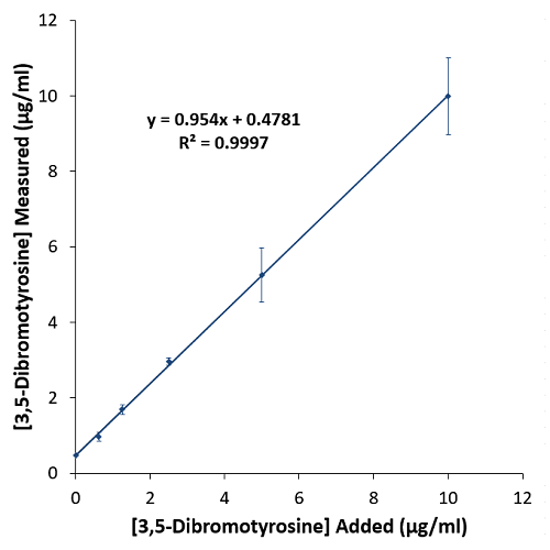
Typical Standard Curve for the Dibromo-tyrosine ELISA Kit (Enzyme-Linked Immunosorbent Assay) MOLECULAR SIGNATURE® – SKT-140. Assay Type: Competitive ELISA. Detection Method: Colorimetric Assay. Assay Range: 0.078 – 5 µg/ml.
Chemical Equation of the Bromination of Tyrosine.
Diagram of the Dibromo-tyrosine Competitive ELISA.
Recovery of dibromo-tyrosine from urine. Urine samples were spiked with dibromo-tyrosine, and diluted as described in the Sample Preparation section (page 10) and analyzed using the Dibromo-Tyrosine ELISA kit. The y-intercept corresponds to the amount of dibromo-tyrosine in unspiked urine.
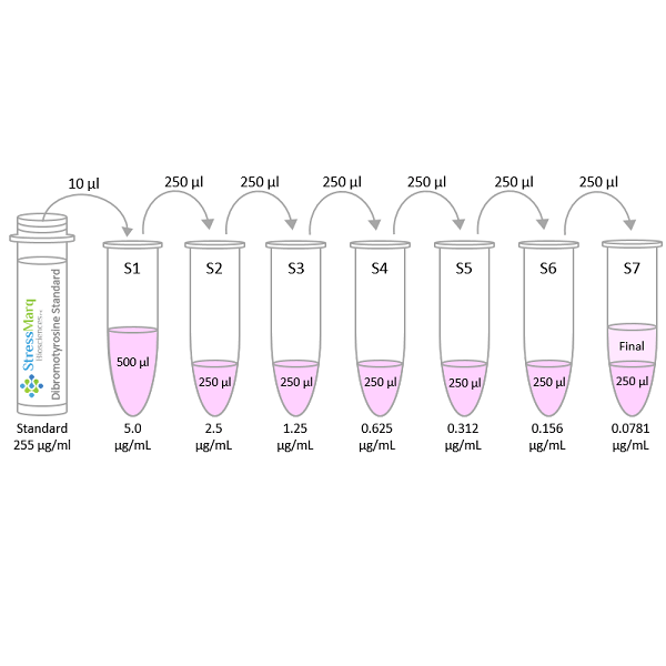
Diagram of the Preparation of the Dibromo-tyrosine Standards.
Diagram of the Triplicate Sample Plate Format.
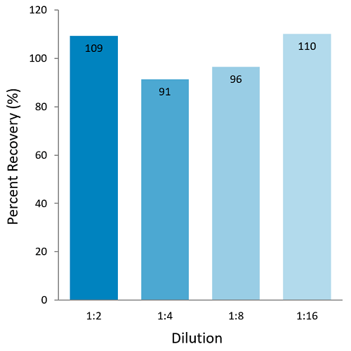
Graph of the Dibromo-tyrosine Spike Recovery in Human Plasma.
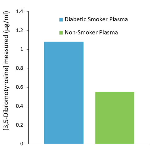
Graph of the sample comparison between diabetic smoker plasma and non-diabetic/non-smoker plasma.
 Powered by Bioz
Reviews
There are no reviews yet.