Discovery through Partnership | Excellence through Quality
| Product Name | Alpha Synuclein Pre-formed Fibrils | |||||||||||||||||||||||||||||||||||||||||||||||||||||||||||||||||||||||||||||||||
| Description |
Human Recombinant Alpha Synuclein PFFs (Type 1) |
|||||||||||||||||||||||||||||||||||||||||||||||||||||||||||||||||||||||||||||||||
| Applications | WB, SDS-PAGE, In vivo assay, In vitro assay | |||||||||||||||||||||||||||||||||||||||||||||||||||||||||||||||||||||||||||||||||
| Concentration | 2 mg/ml or 5 mg/ml | |||||||||||||||||||||||||||||||||||||||||||||||||||||||||||||||||||||||||||||||||
| Conjugates |
No tag
StreptavidinProperties:
Biotin
|
|||||||||||||||||||||||||||||||||||||||||||||||||||||||||||||||||||||||||||||||||
| Nature | Recombinant | |||||||||||||||||||||||||||||||||||||||||||||||||||||||||||||||||||||||||||||||||
| Species | Human | |||||||||||||||||||||||||||||||||||||||||||||||||||||||||||||||||||||||||||||||||
| Expression System | E. coli | |||||||||||||||||||||||||||||||||||||||||||||||||||||||||||||||||||||||||||||||||
| Amino Acid Sequence | MDVFMKGLSK AKEGVVAAAE KTKQGVAEAA GKTKEGVLYV GSKTKEGVVH GVATVAEKTK EQVTNVGGAV VTGVTAVAQK TVEGAGSIAA ATGFVKKDQL GKNEEGAPQE GILEDMPVDP DNEAYEMPSE EGYQDYEPEA | |||||||||||||||||||||||||||||||||||||||||||||||||||||||||||||||||||||||||||||||||
| Purity | >95% | |||||||||||||||||||||||||||||||||||||||||||||||||||||||||||||||||||||||||||||||||
| Other Resources | Sonication Protocol | |||||||||||||||||||||||||||||||||||||||||||||||||||||||||||||||||||||||||||||||||
| Protein Length | Full Length | |||||||||||||||||||||||||||||||||||||||||||||||||||||||||||||||||||||||||||||||||
| Protein Size | ~14.46 kDa | |||||||||||||||||||||||||||||||||||||||||||||||||||||||||||||||||||||||||||||||||
| Biological Activity | Endogenous alpha-synuclein phosphorylation. 100 µM alpha synuclein protein monomer (SPR-321) seeded with 10 uM alpha synuclein protein PFF (SPR-322) in 25 µM Thioflavin T (PBS pH 7.4, 100 µl reaction volume) generated a fluorescence intensity of 13,000 Relative Fluorescence Units after incubation at 37°C with shaking at 600 rpm. Fluorescence was measured by excitation at 450 nm and emission at 485 nm on a Molecular Devices Gemini XPS microplate reader. | |||||||||||||||||||||||||||||||||||||||||||||||||||||||||||||||||||||||||||||||||
| Field of Use | Not for use in humans. Not for use in diagnostics or therapeutics. For in vitro research use only. | |||||||||||||||||||||||||||||||||||||||||||||||||||||||||||||||||||||||||||||||||


Properties
| Storage Buffer | PBS |
| Storage Temperature | -80ºC |
| Shipping Temperature | Dry Ice. Shipping note: Product will be shipped separately from other products purchased in the same order. |
| Purification | Ion-exchange Purified |
| Cite This Product | Human Recombinant Alpha Synuclein Pre-formed Fibrils (StressMarq Biosciences | Victoria, BC CANADA | Catalog# SPR-322) |
| Certificate of Analysis | Certified >95% pure using SDS-PAGE analysis. Low endotoxin <5 EU/mL @ 2mg/mL. |
| Other Relevant Information | For best results, sonicate immediately prior to use. Refer to the Neurodegenerative Protein Handling Instructions on our website, or the product datasheet for further information. Monomer source is catalog# SPR-321. |
Biological Description
| Alternative Names | Alpha-synuclein, Alpha synuclein, Asyn, SNCA, NACP, PARK1, PARK4, PD1, Synuclein alpha, Non-A beta component of AD amyloid, Non-A4 component of amyloid precursor, Synuclein Alpha-140, SYN, Parkinson's disease familial 1 Protein Protein, Alpha Synuclein PFFs |
| Research Areas | Alzheimer's Disease, Neurodegeneration, Neuroscience, Parkinson's Disease, Synuclein, Tangles & Tau, Multiple System Atrophy |
| Cellular Localization | Cytoplasm, Membrane, Nucleus |
| Accession Number | NP_000336.1 |
| Gene ID | 6622 |
| Swiss Prot | P37840 |
| Scientific Background |
Alpha-synuclein (α-syn), a neuronal protein encoded by the SNCA gene, plays a critical role in synaptic function, including vesicle trafficking and neurotransmitter release. Under physiological conditions, α-syn exists primarily in monomeric or tetrameric forms. However, pathological misfolding of α-syn can lead to the formation of pre-formed fibrils (PFFs), which are central to the progression of synucleinopathies such as Parkinson’s disease, Lewy body dementia, and multiple system atrophy. PFFs are structurally stable aggregates that act as seeds for the recruitment and conversion of native α-syn into toxic fibrillar forms. This seeding mechanism initiates a prion-like spread of misfolded α-syn across neuronal networks, leading to the accumulation of Lewy bodies and neurites—hallmarks of neurodegenerative pathology. The presence of PFFs disrupts cellular homeostasis, impairs mitochondrial function, and activates neuroinflammatory responses, contributing to progressive neuronal dysfunction and death. Experimental models utilizing α-syn PFFs have demonstrated their capacity to replicate disease-like features, including synaptic deficits, motor impairments, and widespread protein aggregation. These models are instrumental in elucidating disease mechanisms and identifying therapeutic targets. Targeting the formation, propagation, or cellular uptake of α-syn PFFs represents a promising strategy for disease modification. Advances in understanding the molecular behavior of PFFs are paving the way for novel interventions aimed at halting or reversing neurodegenerative processes. |
| References |
1. “Genetics Home Reference: SNCA”. US National Library of Medicine. (2013). 2. Zhang L., et al. (2008) Brain Res. 1244: 40-52. 3. Alim M.A., et al. (2002) J Biol Chem. 277(3): 2112-2117. 4. Kokhan V.S., Afanasyeva M.A., Van'kin G. (2012) Behav. Brain. Res. 231(1): 226-230. 5. Spillantini M.G., et al. (1997) Nature. 388(6645): 839-840. 6. Mezey E., et al. (1998) Nat Med. 4(7): 755-757. |
Product Images
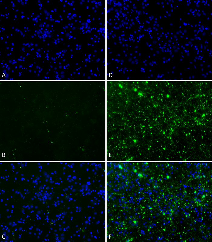
Primary rat hippocampal neurons show lewy body inclusion formation when treated with Type 1 Alpha Synuclein Protein Pre-formed Fibrils (SPR-322) at 4 µg/ml (D-F), but not when treated with Type 2 Alpha Synuclein Protein Pre-formed Fibrils (SPR-317) at 4 µg/ml (A-C). Tissue: Primary hippocampal neurons. Species: Sprague-Dawley rat. Fixation: 4% formaldehyde from PFA. Primary Antibody: Mouse anti-pSer129 Antibody at 1:1000 24 hours at 4°C. Secondary Antibody: FITC Goat Anti-Mouse (green) at 1:700 for 1 hours at RT. Counterstain: Hoechst (blue) nuclear stain at 1:4000 for 1 hour at RT. Localization: Lewy body inclusions. Magnification: 20x.
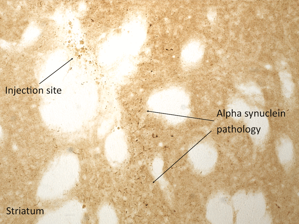
Immunohistochemistry analysis of rat brain injected with Type 1 human alpha synuclein PFFs (SPR-322). Species: Female Sprague-Dawley Rat. Rat was injected with 16µg Type 1 human alpha synuclein PFFs (SPR-322) in each of 2 injection sites: AP+1.6, ML+2.4, DV-4.2 from skull; and AP-1.4, ML+0.2, DV-2.8 from skull. 30-days post-injection. Fixation: Saline perfusion followed by 4% PFA fixation for 48 hrs. Primary antibody: rabbit monoclonal anti-pSer129 alpha synuclein. Secondary Antibody: Biotin-SP Donkey Anti-Rabbit IgG (H+L) at 1:500 for 2 hours in cold room with shaking. ABC signal amplification, DAB staining. Magnification: 20X. Alpha synuclein pathology is seen in the striatum close to an injection site.
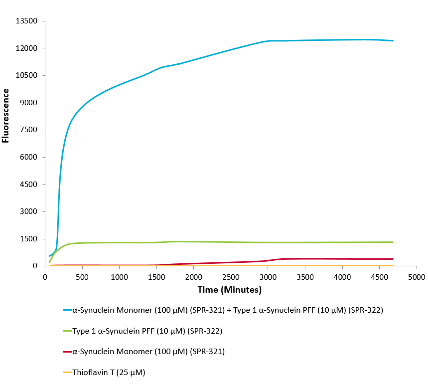
Type 1 alpha synuclein Pre-formed fibrils (SPR-322) seed the formation of new alpha synuclein fibrils from the pool of alpha synuclein monomers (SPR-321). Thioflavin T is a fluorescent dye that binds to beta sheet-rich structures, such as those in alpha synuclein fibrils. Upon binding, the emission spectrum of the dye experiences a red-shift, and increased fluorescence intensity. Thioflavin T emission curves show increased fluorescence (correlated to alpha synuclein protein aggregation) over time when 10 µM of Type 1 alpha synuclein Pre-formed fibrils (SPR-322) is combined with 100 µM of alpha synuclein monomer (SPR-321), as compared to Type 1 alpha synuclein Pre-formed fibrils (SPR-322) alone and alpha synuclein monomer (SPR-321) alone. Thioflavin T ex = 450 nm, em = 485 nm. Note: We use molecular weight of 14.46 kDa for both alpha synuclein monomer and fibril in calculations. We load 100µL/well for Thioflavin T assay so 100 µM is 144.6µg/well and 10 µM is 14.46 µg/well.
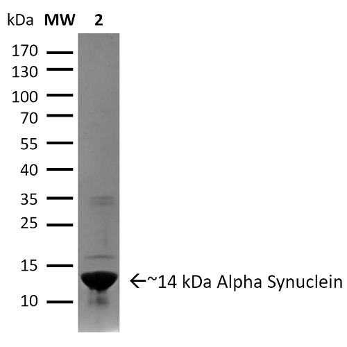
SDS-PAGE of ~14 kDa Type 1 Human Recombinant Alpha Synuclein Protein Pre-formed Fibrils (SPR-322). Lane 1: Molecular Weight Ladder (MW). Lane 2: Type 1 Alpha Synuclein Protein Pre-formed Fibrils (SPR-322).
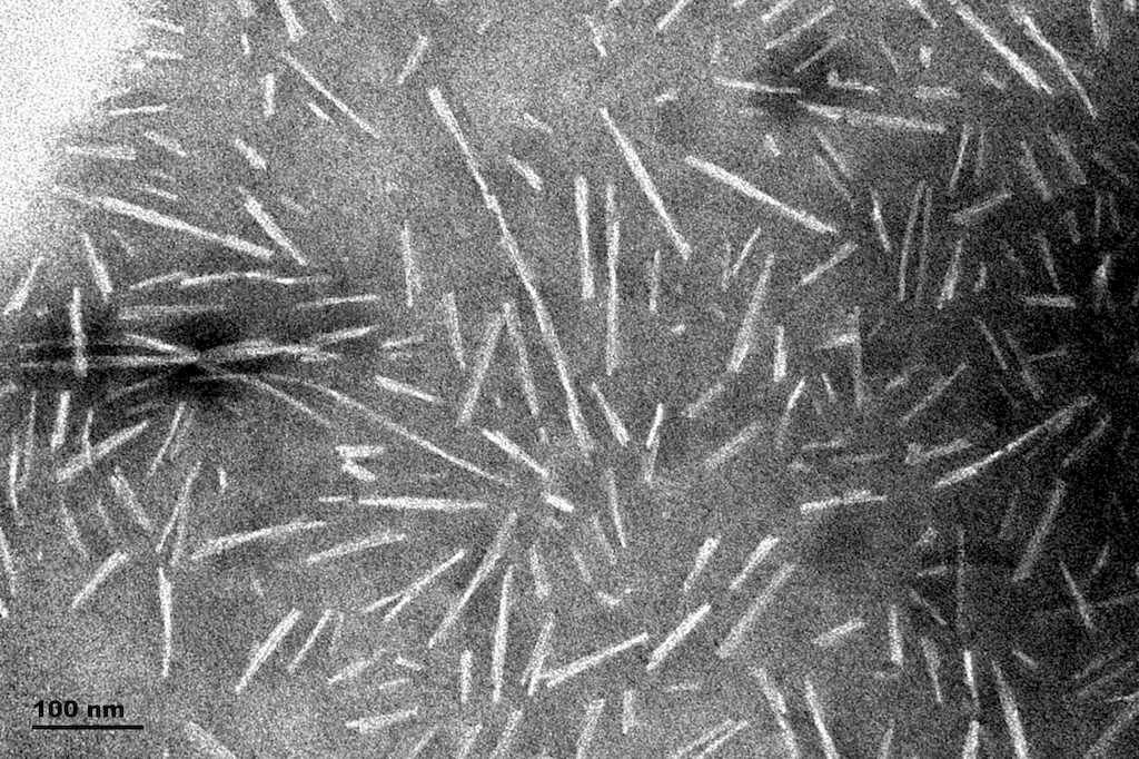
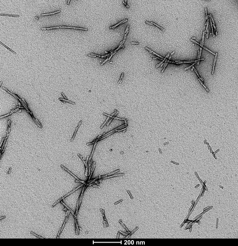
TEM of Type 1 Alpha Synuclein Pre-formed Fibrils (PFFs) (SPR-322)
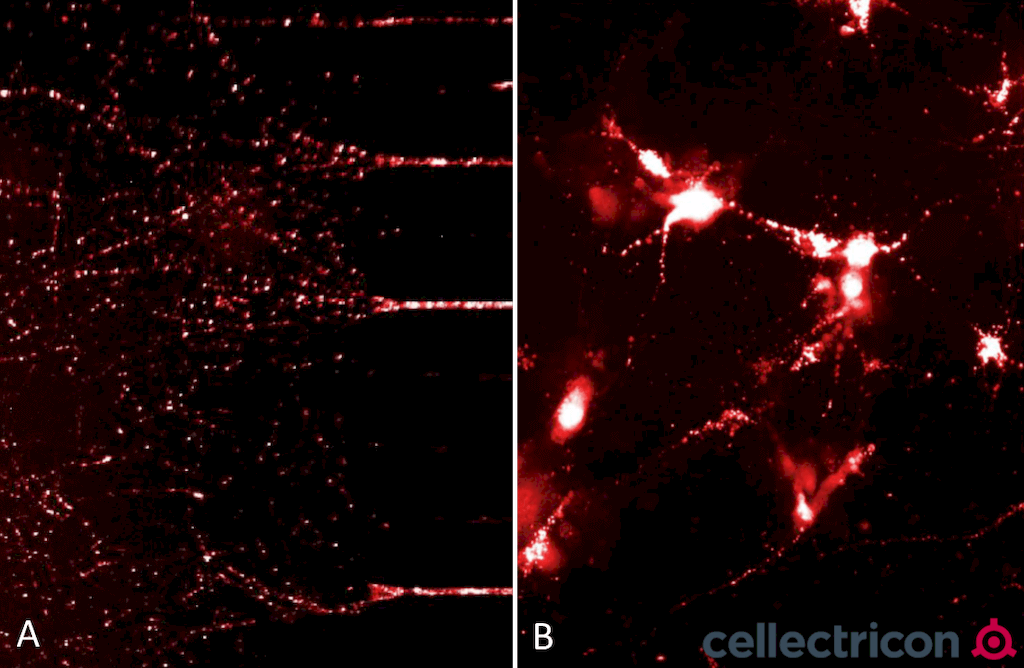
ATTO633 fluorescently-labelled alpha synuclein PFFs (SPR-322) were taken up, transported into the soma, and induced alpha synuclein aggregation in mouse neurocortical primary cells. (A) Neurites filled with fluorescently-labelled alpha synuclein seeds in a microfluidic co-culture system after 24 hours. (B) Alpha synuclein seeds within the soma and neurites of mouse neurocortical primary cells after 24 hours. Experiment and imaging courtesy of Cellectricon.
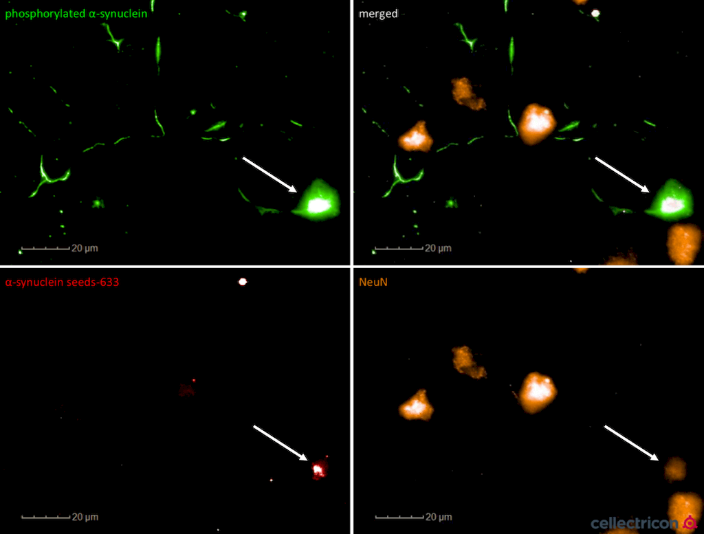
Confocal imaging shows NeuN+ (mature) primary cortical neurons filled with ATTO633 fluorescently-labelled alpha synuclein PFFs (SPR-322). ATTO-633 alpha synuclein PFFs seed endogenous alpha synculein phosphorylation after 7 days. Experiment and imaging courtesy of Cellectricon.
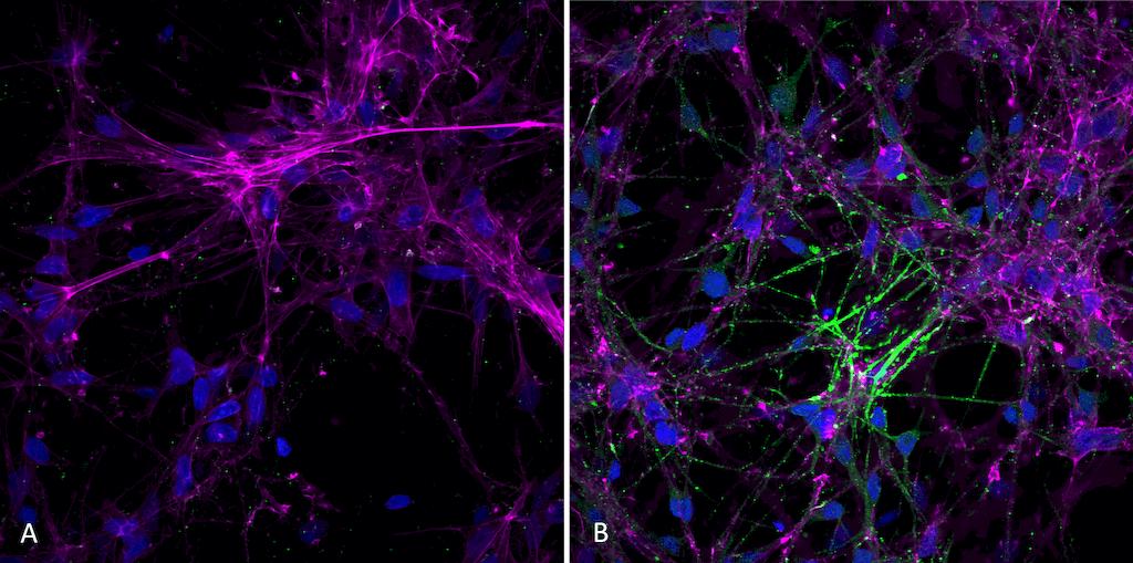
Immunocytochemistry/Immunofluorescence analysis of human iPSC-derived neurons treated with alpha synuclein pre-formed fibrils (SPR-322). Primary Antibody: Mouse Anti-Alpha Synuclein (pSer129) Monoclonal Antibody (SMC-600) at 1:1000 for O/N at 4°C. Secondary Antibody: Anti-Rabbit: A488 at 1:1000 for 1 hour at RT. Magnification: 40X. Nuclear stain: Hoechst- 20 min, RT (blue). Actin stain: Phalloidin-647- 20 min, RT (magenta). 4K cells per well. iPSC neurons: Applied StemCell Catalog # ASE-9321K. A) negative control; no fibrils added to well. B) 7 days after addition of active recombinant human pre-formed fibrils (Type 1). Fibrils were sonicated before use and applied 2.5 ug per well
TEM of Type 1 Alpha Synuclein Pre-formed Fibrils (PFFs) (SPR-322)
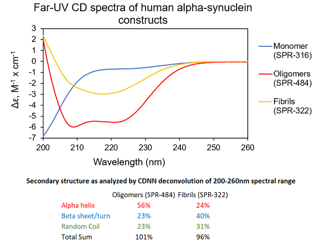
Αlpha Synuclein Oligomers Have Distinct Secondary Structure Differences Compared to Fibrils. UV-CD data suggests that StressMarq’s Alpha Synuclein Oligomers have distinct secondary structure differences compared to our monomers and fibrils. More specifically, StressMarq’s Kinetically Stable Alpha Synuclein Oligomers (SPR-484) show a significantly higher alpha helix content and lower beta sheet/turn content than our Alpha Synuclein Pre-formed Fibrils (Type 1) (SPR-322). StressMarq’s Alpha Synuclein Monomers (SPR-316) show a strong negative signal at 200 nm indicative of a disordered protein state (low secondary structure content).
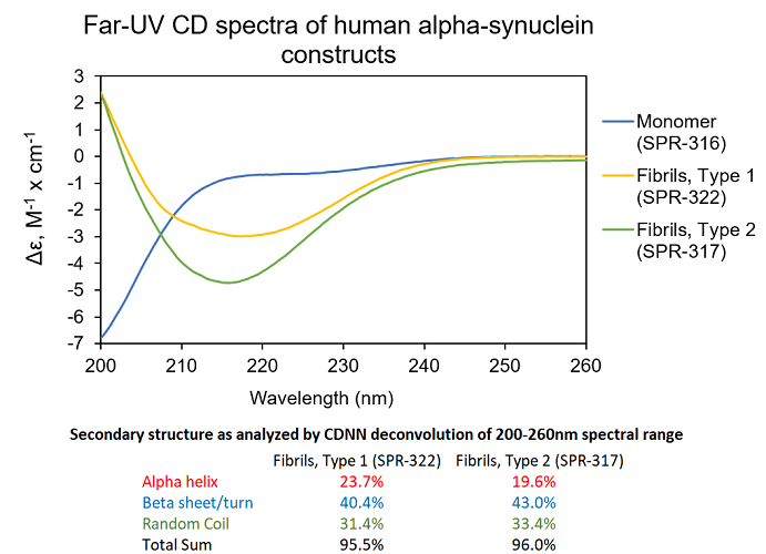
UV-CD data suggests StressMarq’s Alpha Synuclein Pre-formed Fibrils (PFF), Type 1 (cat#SPR-322) and Type 2 (cat#SPR-317) both have a high beta sheet/turn content, yet do have small secondary structure differences. StressMarq’s Alpha Synuclein Monomers (cat#SPR-316) show a strong negative signal at 200 nm indicative of a disordered protein state (low secondary structure content). For this experiment, pre-formed fibrils (PFF) were subjected to 10 cycles of sonication prior to UV-CD to ensure solubility prior to measurement.
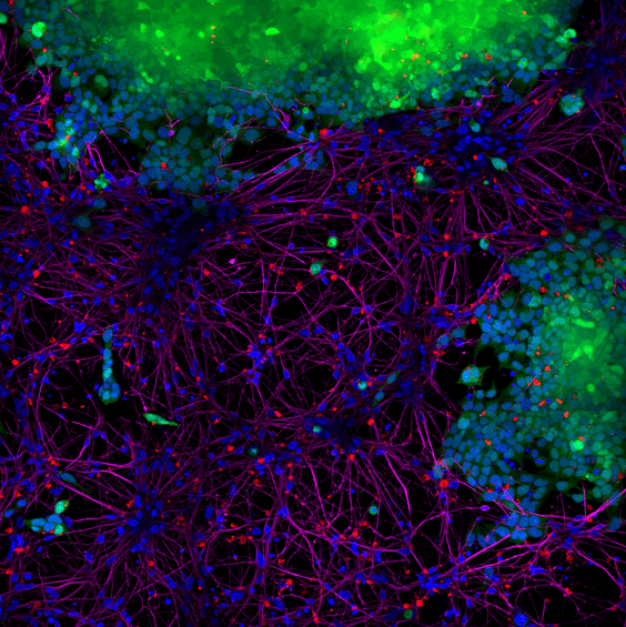
StressMarq’s Alpha Synuclein PFFs (red) cat#SPR-322, were shown to be taken up by SH-SY5Y cells and transmitted to neuronal iPSCs within 14 days. Blue: Hoechst/DNA; Green: SHSY5Y-GFP; Red: alpha-synuclein PFFs-555 (cat#SPR-322); Purple: Tubulin.
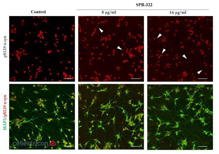
Representative fluorescence images on human iPSC-derived neuronal cultures at DIV 35 (PFFs added at DIV 7, followed by a 4-week incubation). SPR-322 was added at either 8 µg/mL or 16 µg/mL, with the control having none added. Anti-pS129 (red) was used to highlight alpha synuclein aggregation features, such as larger puncta and beads-on-a-string (white arrowheads) not seen in the control. Data courtesy of Cellectricon.
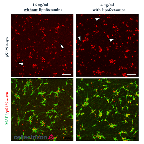
Representative fluorescence images on human iPSC-derived neuronal cultures at DIV 28 (PFFs added at DIV 14, followed by a 2-week incubation). SPR-322 was added at either 4 µg/mL with lipofectamine or 16 µg/mL without lipofectamine. Anti-pS129 (red) was used to highlight alpha synuclein aggregation features, such as larger puncta and beads-on-a-string (white arrowheads). Data courtesy of Cellectricon.
 Powered by Bioz
StressMarq Biosciences :
Based on validation through cited publications.