Discovery through Partnership | Excellence through Quality
| Product Name | FCR1 |
| Description |
FRET ratiometric fluorescence-based redox sensor |
| Molecular Formula | C34H37N7O6 |
| Molecular Weight | 639 |
| Field of Use | Not for use in humans. Not for use in diagnostics or therapeutics. For in vitro research use only. |
Properties
| Storage Temperature | -20ºC |
| Shipping Temperature | Blue Ice or 4ºC |
| Product Type | Redox Probe |
| Solubility | Soluble in DMSO |
| Source | Synthetic |
| Appearance | Orange Solid |
| Safety Phrases |
Classification: Caution: Substance not yet fully tested. Safety Phrases: S22 - Do not breathe dust S24/25 - Avoid contact with skin and eyes S36/37/39 - Wear suitable protective clothing, gloves and eye/face protection |
| Cite This Product | FCR1 (StressMarq Biosciences Inc., Victoria BC CANADA, Catalog # SIH-180) |
Biological Description
| Alternative Names | 7-(Diethylamino)-N-((1r,4r)-4-(2-(10-ethyl-2,4-dioxo-4,10-dihydrobenzo[g]pteridin-3(2H)-yl)acetamido)cyclohexyl)-2-oxo-2H-chromene-3-carboxamide (FCR1) |
| Research Areas | Cancer, Oxidative Stress |
| Scientific Background | FCR1 (Flavin Coumarin Redox Sensor 1) is a ratiometric fluorescent probe designed to monitor cellular redox states with high sensitivity. In neuroscience, FCR1 enables real-time visualization of oxidative stress dynamics in neural tissues using fluorescence lifetime imaging microscopy and flow cytometry. Its reversible fluorescence shift upon redox changes allows researchers to track oxidative capacity in neurons and glial cells without interference from background signals. This makes FCR1 a valuable tool for studying neurodegenerative diseases where redox imbalance plays a critical role. |
| References | 1. Kaur A., Haghighatbin M.A., Hogan C.F., and New E.J. (2015) Chem. Commun. Epub. |
Product Images
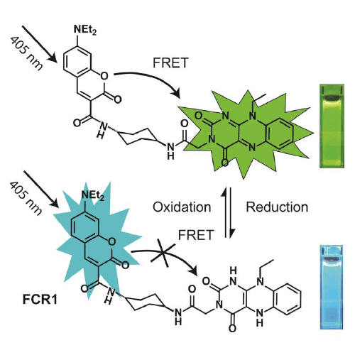
Chemical structure and design of FCR1 (SIH-180), showing FRET processes in oxidised form. Inset: photographs of cuvettes of FCR1 in oxidised and reduced forms under 365 nm excitation. Images used with permission from Kaur A, Haghighatbin MA, Hogan CF, New EJ. Chem Commun (Camb). 2015 Jun 16;51(52):10510-3.
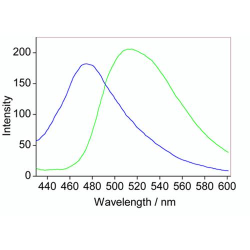
Fluorescence behavior of FCR1 (SIH-180) in the oxidized (green) and reduced (blue) forms, using 10 µM. Excitation: 405 nm. Reduced Emission: 475 nm. Oxidized Emission: 520 nm. Images used with permission from Kaur A, Haghighatbin MA, Hogan CF, New EJ. Chem Commun (Camb). 2015 Jun 16;51(52):10510-3.
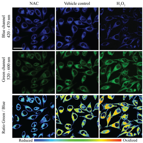
Two photon – confocal microscopy imaging of HeLa cells treated with FCR1 (SIH-180, 10 µM, 15 min, λex = 820 nm) and (a) N-acetyl cysteine (50 µM, 30 min), (b) vehicle control and (c) H2O2 (50 µM, 30 min) in blue and green channels. The pseudo colour ratio images indicate the ratio of emission intensity in the green channel to blue channel. Scale bar represents 20 µm. Images used with permission from Kaur A, Haghighatbin MA, Hogan CF, New EJ. Chem Commun (Camb). 2015 Jun 16;51(52):10510-3.
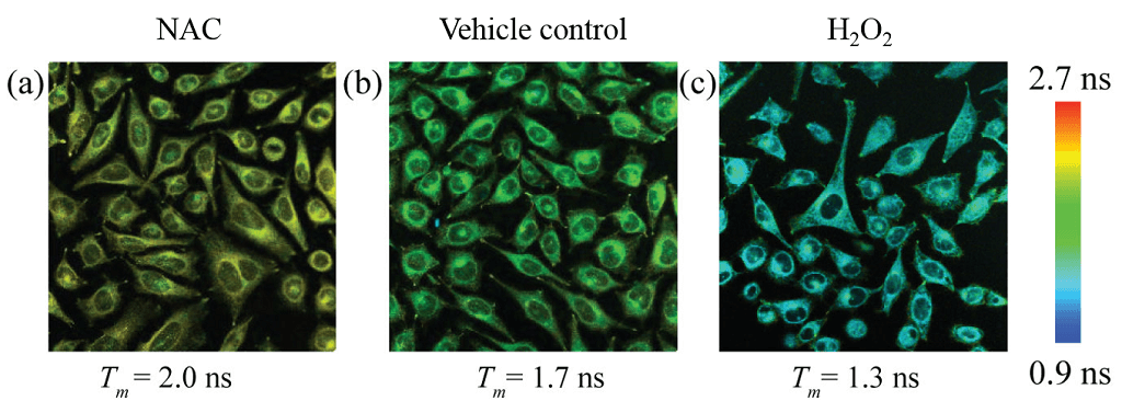
Fluorescence lifetimes of the donor (420 – 470 nm) in HeLa cells treated with FCR1 (SIH-180, 10 µM, λex = 820 nm) and N-acetyl cysteine, vehicle control and H2O2. Pseudo-colour images represent mean lifetime. Scale bar represents 50 µm. Images used with permission from Kaur A, Haghighatbin MA, Hogan CF, New EJ. Chem Commun (Camb). 2015 Jun 16;51(52):10510-3.
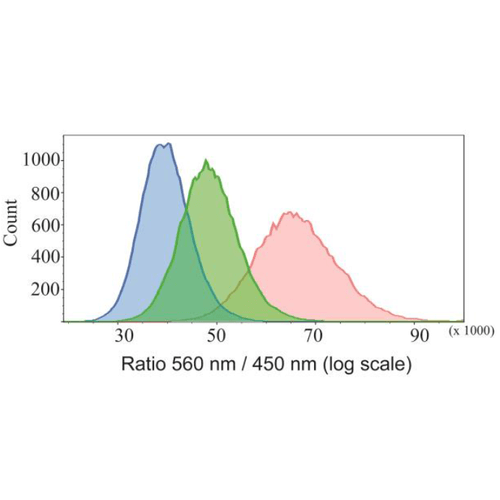
Flow cytometric studies of HeLa cells treated with FCR1 (SIH-180, 10 µM, λex = 405 nm) showing the fluorescence ratio (560 / 450 nm) when treated with N-acetyl cysteine (blue), vehicle control (green) and H2O2 (red). Images used with permission from Kaur A, Haghighatbin MA, Hogan CF, New EJ. Chem Commun (Camb). 2015 Jun 16;51(52):10510-3.
 Powered by Bioz
Reviews
There are no reviews yet.