Discovery through Partnership | Excellence through Quality
| Product Name | NpFR1 |
| Description |
Reversible fluorescence intensity-based redox sensor |
| Molecular Formula | C23H21N5O4 |
| Molecular Weight | 431.45 |
| Field of Use | Not for use in humans. Not for use in diagnostics or therapeutics. For in vitro research use only. |
Properties
| Storage Temperature | -20ºC |
| Shipping Temperature | Blue Ice or 4ºC |
| Product Type | Redox Probe |
| Solubility | Soluble in DMSO |
| Source | Synthetic |
| Appearance | Red Solid |
| SMILES | C1(N(C(C2=CC5=C(C3=CC=CC1=C23)N(C4=NC(NC(C4=N5)=O)=O)CCC)=O)CCCC)=O |
| InChI | InChI=1S/C23H21N5O4/c1-3-5-10-28-21(30)13-8-6-7-12-16(13)14(22(28)31)11-15-18(12)27(9-4-2)19-17(24-15)20(29)26-23(3 |
| InChIKey | C5(C2=C1C(=CC4=C(C1=CC=C2)N(C3=NC(NC(C3=N4)=O)=O)CCC)C(N5CCCC)=O)=O |
| Safety Phrases |
Classification: Caution: Substance not yet fully tested. Safety Phrases: S22 - Do not breathe dust S24/25 - Avoid contact with skin and eyes S36/37/39 - Wear suitable protective clothing, gloves and eye/face protection |
| Cite This Product | NpFR1 (StressMarq Biosciences Inc., Victoria BC CANADA, Catalog # SIH-181) |
Biological Description
| Research Areas | Cancer, Oxidative Stress |
| Scientific Background | NpFR1 (Napthalimide-Flavin Redox Sensor 1) is a highly sensitive fluorescent probe that exhibits a dramatic increase in fluorescence upon oxidation. Its reversible redox behavior allows for dynamic monitoring of oxidative stress in live cells. In neuroscience, NpFR1 is particularly useful for detecting redox fluctuations in neuronal environments, aiding in the study of neurodegenerative conditions such as ALS and Parkinson’s disease. Its high signal-to-noise ratio and responsiveness to mild oxidants make it ideal for tracking oxidative damage in neural tissues. |
| References | 1. Yeow J., Kaur A.K., Anscomb M.D., and New E.J. (2014) Chem. Commun. 50: 8181-8184. |
Product Images
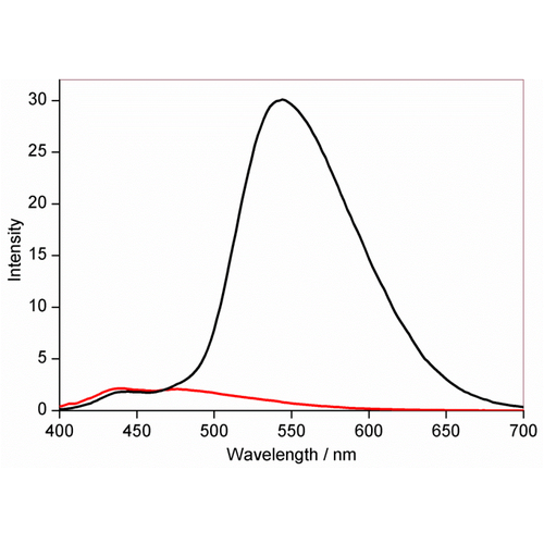
Fluorescence behavior of NpFR1 (SIH-181) in the oxidized (black) and reduced (red) forms, using 50 µM. Excitation: 405 or 488 nm (not shown). Emission: 490 – 600 nm, with peak at 545 nm.
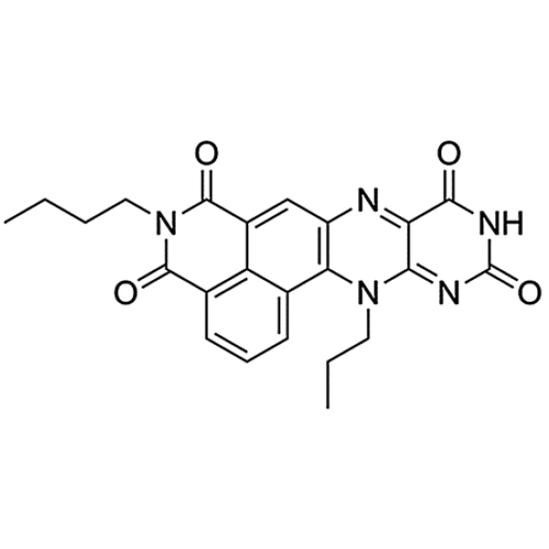
Chemical structure of NpFR1 (SIH-181), a reversible fluorescence intensity-based redox sensor. This image is from Chem. Commun., 2014, 50, 8181, and licensed under a Creative Commons Attribution 3.0 Unported Licence (http://creativecommons.org/licenses/by/3.0/).
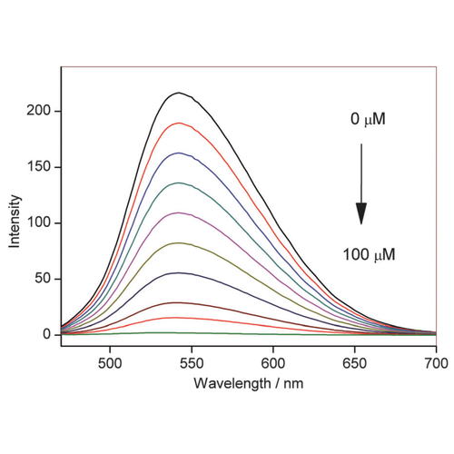
Fluorescence emission of NpFR1 (SIH-181, 5 mM, lex = 405 nm) with the incremental addition of sodium dithionite. All spectra were acquired in HEPES buffer (100 mM, pH 7.4). This image is from Chem. Commun., 2014, 50, 8181, and licensed under a Creative Commons Attribution 3.0 Unported Licence (http://creativecommons.org/licenses/by/3.0/).
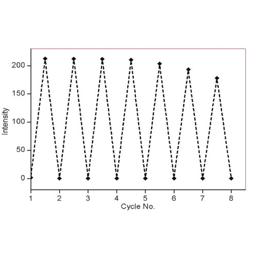
Fluorescence response of NpFR1 (SIH-181) to cycles of oxidation and reduction. Reduction was achieved with sodium dithionite (100mM) followed by re-oxidation with 250mM H2O2. Spectra were recorded 5 min after the addition of reducing and oxidising agents. All spectra were acquired in HEPES buffer (100 mM, pH 7.4). This image is from Chem. Commun., 2014, 50, 8181, and licensed under a Creative Commons Attribution 3.0 Unported Licence (http://creativecommons.org/licenses/by/3.0/).
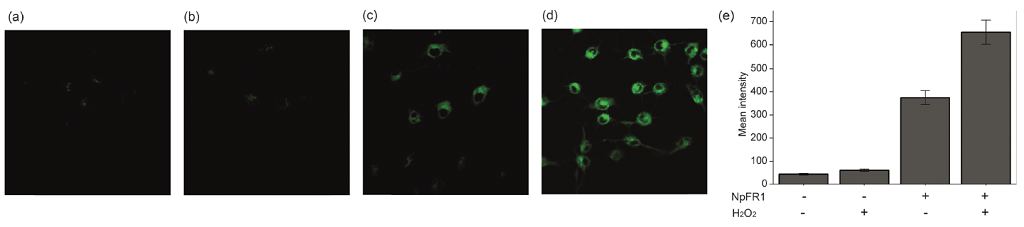
Imaging of NpFR1 (SIH-181) in 3T3-L1 preadipocytes treated with (a) vehicle control (b) H2O2 (100 mM, 2 min), (c) NpFR1 (50 mM, 2 h) and (d) NpFR1 (50 mM, 2 h) followed by H2O2 (100 mM, 2 min). Scale bar represents 50 mm, lex = 405 nm. (e) Integrated emission from 510 nm to 610 nm. Values are the mean ratio generated from the intensity from five fields of cells. Error bars represent standard error measurement (s.e.m.). The layout of this image was modified for optimal display from the original Chem. Commun., 2014, 50, 8181, and licensed under a Creative Commons Attribution 3.0 Unported Licence (http://creativecommons.org/licenses/by/3.0/).
 Powered by Bioz
Reviews
There are no reviews yet.