Advancing the Frontiers of Neurodegenerative Disease Research


Properties
| Storage Buffer | PBS pH7.2, 50% glycerol, 0.09% sodium azide *Storage buffer may change when conjugated |
| Storage Temperature | -20ºC, Conjugated antibodies should be stored according to the product label |
| Shipping Temperature | Blue Ice or 4ºC |
| Purification | Protein G Purified |
| Clonality | Monoclonal |
| Clone Number | H9010 |
| Isotype | IgG2a |
| Specificity | Detects 90kDa. Detects HSP90 beta in all reactive species except in Chicken, where it detects both alpha and beta isoforms. |
| Cite This Product | HSP90 Antibody (StressMarq Biosciences | Victoria, BC CANADA, Catalog# SMC-107, RRID: AB_854214) |
| Certificate of Analysis | 1 µg/ml of SMC-107 was sufficient for detection of HSP90beta in 20 µg of heat shocked HeLa cell lysate by colorimetric immunoblot analysis using Goat anti-mouse IgG:HRP as the secondary antibody. |
Biological Description
| Alternative Names | HSP90, HSP90AB1, HSP90-beta, HSPCB, HSPC2, Heat shock protein HSP 90-beta, Heat shock 84 kDa protein, HSP84, HSP90B |
| Research Areas | Cancer, Cell Signaling, Chaperone Proteins, Heat Shock, Protein Trafficking, Tumor Biomarkers |
| Cellular Localization | Cytoplasm, Melanosome |
| Accession Number | NP_031381.2 |
| Gene ID | 3326 |
| Swiss Prot | P08238 |
| Scientific Background |
HSP90 is a highly conserved and abundantly expressed molecular chaperone that plays a central role in maintaining protein homeostasis, particularly in the nervous system. Present in all eukaryotic cells, HSP90 exists in two major cytosolic isoforms—HSP90α and HSP90β—which share 85% sequence identity but differ in oligomeric state and regulatory function. Despite its classification as a heat shock protein, HSP90 is constitutively expressed at high levels, comprising up to 2% of total cytosolic protein in unstressed cells. It is essential for the folding, maturation, and stabilization of a wide range of client proteins, many of which are involved in neuronal signaling, synaptic plasticity, and stress response. These include kinases (e.g., c-Raf), transcription factors (e.g., p53), and steroid hormone receptors. In neurodegenerative diseases such as Alzheimer’s, Parkinson’s, and Huntington’s, HSP90 is implicated in both protective and pathological processes. It stabilizes misfolded proteins and prevents aggregation, but can also shield aberrant proteins from degradation, contributing to disease progression. HSP90’s interaction with co-chaperones like Cdc37 and p23 forms complexes that regulate the fate of client proteins, making it a key node in proteostasis networks. Pharmacological inhibition of HSP90—using compounds like geldanamycin—has shown promise in modulating protein quality control pathways and reducing toxic protein accumulation in neurodegenerative models. |
| References |
1. Nemoto T., et al. (1997) J.Biol Chem. 272: 26179-26187. 2. Minami Y., et al. (1991), J.Biol Chem. 266: 10099-10103. 3. Arlander S.J.H., et al. (2003) J Biol Chem 278: 52572-52577. 4. Pearl H., et al. (2001) Adv Protein Chem 59:157-186. 5. Neckers L., et al. (2002) Trends Mol Med 8:S55-S61. 6. Pratt W., Toft D. (2003) Exp Biol Med 228:111-133. 7. Pratt W., Toft D. (1997) Endocr Rev 18:306–360. 8. Pratt W.B. (1998) Proc Soc Exptl Biol Med 217: 420–434. 9. Whitesell L., et al. (1994) Proc Natl Acad Sci USA 91: 8324–8328. 10. Barent R. L. (1998) Mol. Endocrinol. 12: 342-354 11. Lo. M.A. (1998) EMBO J. 17: 6879-6887. |
Product Images
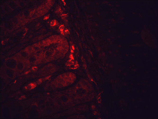
Immunohistochemistry analysis using Mouse Anti-Hsp90 Monoclonal Antibody, Clone H9010 (SMC-107). Tissue: inflamed colon. Species: Mouse. Fixation: Formalin. Primary Antibody: Mouse Anti-Hsp90 Monoclonal Antibody (SMC-107) at 1:10000 for 12 hours at 4°C. Secondary Antibody: Alexa Fluor 555 Goat Anti-Mouse (red) at 1:5000 for 1 hour at RT. Localization: Inflammatory and epithelial mucosa. Magnification: 40x. Inflammatory and epithelial mucosa. This image was produced using an amplifying IHC wash buffer. The antibody has therefore been diluted more than is recommended for other applications.
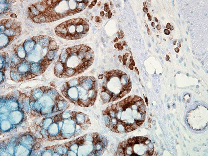
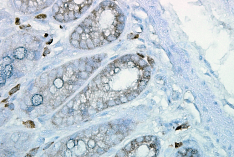
Immunohistochemistry analysis using Mouse Anti-Hsp90 Monoclonal Antibody, Clone H9010 (SMC-107). Tissue: colon carcinoma. Species: Human. Fixation: Formalin. Primary Antibody: Mouse Anti-Hsp90 Monoclonal Antibody (SMC-107) at 1:10000 for 12 hours at 4°C. Secondary Antibody: Biotin Goat Anti-Mouse at 1:2000 for 1 hour at RT. Counterstain: Mayer Hematoxylin (purple/blue) nuclear stain at 200 µl for 2 minutes at RT. Localization: Inflammatory cells. Magnification: 40x. This image was produced using an amplifying IHC wash buffer. The antibody has therefore been diluted more than is recommended for other applications.
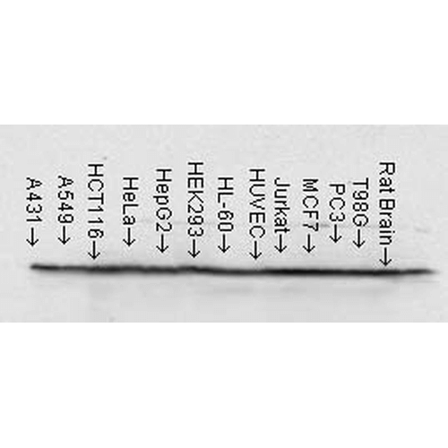
Western Blot analysis of Human cell lysates from various cell lines showing detection of Hsp90 protein using Mouse Anti-Hsp90 Monoclonal Antibody, Clone H9010 (SMC-107). Load: 15 µg. Block: 1.5% BSA for 30 minutes at RT. Primary Antibody: Mouse Anti-Hsp90 Monoclonal Antibody (SMC-107) at 1:1000 for 2 hours at RT. Secondary Antibody: Sheep Anti-Mouse IgG: HRP for 1 hour at RT.
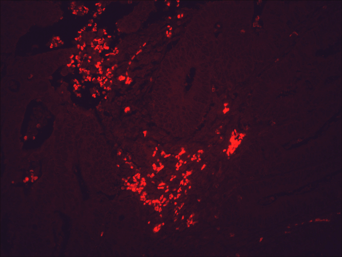
Immunohistochemistry analysis using Mouse Anti-Hsp90 Monoclonal Antibody, Clone H9010 (SMC-107). Tissue: colon carcinoma. Species: Human. Fixation: Formalin. Primary Antibody: Mouse Anti-Hsp90 Monoclonal Antibody (SMC-107) at 1:10000 for 12 hours at 4°C. Secondary Antibody: Alexa Fluor 555 Goat Anti-Mouse (red) at 1:5000 for 1 hour at RT. Magnification: 40x. This image was produced using an amplifying IHC wash buffer. The antibody has therefore been diluted more than is recommended for other applications.
Immunohistochemistry analysis using Mouse Anti-Hsp90 Monoclonal Antibody, Clone H9010 (SMC-107). Tissue: inflamed colon. Species: Mouse. Fixation: Formalin. Primary Antibody: Mouse Anti-Hsp90 Monoclonal Antibody (SMC-107) at 1:10000 for 12 hours at 4°C. Secondary Antibody: Biotin Goat Anti-Mouse at 1:2000 for 1 hour at RT. Counterstain: Mayer Hematoxylin (purple/blue) nuclear stain at 200 µl for 2 minutes at RT. Localization: Inflammatory cells. Magnification: 40x. This image was produced using an amplifying IHC wash buffer. The antibody has therefore been diluted more than is recommended for other applications.
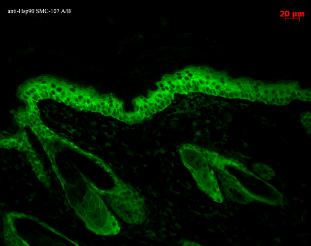
Immunohistochemistry analysis using Mouse Anti-Hsp90 Monoclonal Antibody, Clone H9010 (SMC-107). Tissue: backskin. Species: Mouse. Fixation: Bouin’s Fixative and paraffin-embedded. Primary Antibody: Mouse Anti-Hsp90 Monoclonal Antibody (SMC-107) at 1:100 for 1 hour at RT. Secondary Antibody: FITC Goat Anti-Mouse (green) at 1:50 for 1 hour at RT. Localization: Epidermis.
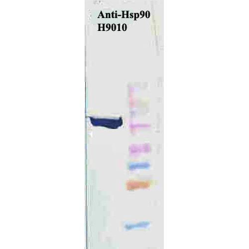
Western Blot analysis of Human Cervical cancer cell line (HeLa) lysate showing detection of Hsp90 protein using Mouse Anti-Hsp90 Monoclonal Antibody, Clone H9010 (SMC-107). Primary Antibody: Mouse Anti-Hsp90 Monoclonal Antibody (SMC-107) at 1:1000. Secondary Antibody: HRP Goat Anti-Mouse.
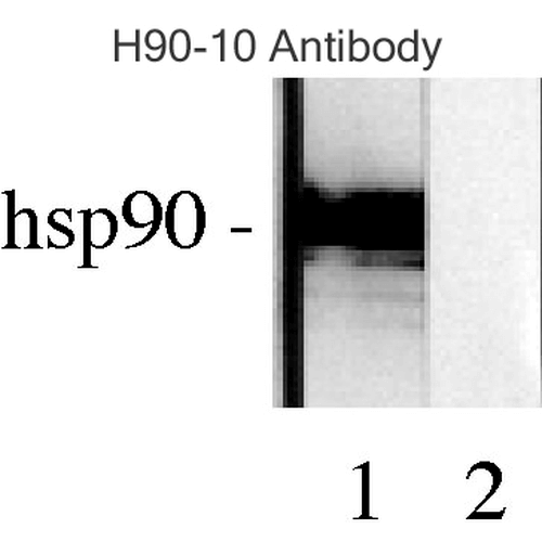
Western blot analysis of Human Lysates showing detection of Hsp90 protein using Mouse Anti-Hsp90 Monoclonal Antibody, Clone H9010 (SMC-107). Primary Antibody: Mouse Anti-Hsp90 Monoclonal Antibody (SMC-107) at 1:1000. Comparison of clone H9010 behavior with Hsp90 human beta (1) and Hsp90 human alpha (2). Courtesy of: David Toft, Mayo Clinic.
 Powered by Bioz
StressMarq Biosciences :
Based on validation through cited publications.