
Optimizing Alpha Synuclein Seed Amplification Assays
Several neurodegenerative diseases are associated with the accumulation of abnormal aggregates of the alpha synuclein protein. These aggregated, pathological forms are the primary components of Lewy bodies (LBs) and Lewy neurites in Parkinson’s disease (PD). In multiple system atrophy (MSA), alpha synuclein aggregates accumulate in oligodendrocytes, forming glial cytoplasmic inclusions (GCIs).
Under healthy physiological conditions, alpha synuclein typically exists as an intrinsically disordered monomer within the cellular cytoplasm. However, in disease states, the protein undergoes a dramatic structural transformation. Its central unstructured region adopts beta-sheet-rich secondary structures, forming oligomers and fibrils. These aggregated forms exhibit prion-like properties, enabling them to seed the misfolding and aggregation of monomeric alpha synuclein, thereby promoting their spread within and between cells.
To detect these pathogenic aggregates, researchers have adapted previously described seed amplification assays (SAAs). This family of assays was originally developed for identifying infectious prion proteins in Creutzfeldt-Jakob disease (CJD). Notable examples include real-time quaking-induced conversion (RT-QuIC) and protein misfolding cyclic amplification (PMCA). Indeed, SAAs have proven instrumental in detecting pathological alpha synuclein in cerebrospinal fluid (CSF) and peripheral tissues, serving as valuable biomarkers for early diagnosis and monitoring of disease progression.
A novel seed amplification assay
Seed amplification assays are powerful diagnostic tools used to detect synucleinopathies, characterized by the accumulation of abnormal alpha synuclein aggregates in neurons. These assays use monomeric alpha synuclein as a substrate. If the sample contains aggregated alpha synuclein, these aggregates act as seeds, triggering the misfolding and aggregation of the monomeric substrate. As the aggregates form beta-sheet-rich fibrils, they bind to Thioflavin T (ThT) dye, which emits a strong fluorescent signal upon excitation—allowing real-time monitoring of the aggregation process.
SAA protocols typically involve repetitive cycles of shaking or sonication followed by incubation. These essential experimental steps fragment aggregates and increase interactions between seeds and monomers. However, sample degradation and the formation of fragments that are too small to act as effective seeds may occur, ultimately reducing assay efficiency.
To address these limitations, Mao et al. developed an improved method known as the Quiescent Seed Amplification Assay (QSAA). This technique eliminates the need for shaking or sonication, relying instead on a temperature-controlled fluorescence reader. The QSAA not only simplifies the SAA protocol but also enhances sensitivity and specificity, achieving over 90% accuracy in distinguishing samples from PD patients and healthy controls. Moreover, this QSAA is versatile, capable of analyzing both brain and skin biopsy samples.
These advancements position the QSAA as a significant improvement over the traditional SAA, offering a more efficient and accessible approach for detecting neurotoxic alpha synuclein aggregates. As a result, it holds great promise for transforming the diagnosis of Parkinson’s disease and related disorders.
Experimental optimization
The research conducted at Guangzhou Medical University, China, aimed to develop an optimized seed amplification assay using quiescent conditions, thereby eliminating the need for mechanical agitation such as shaking or sonication. The study began by identifying the key parameters required to establish an effective SAA protocol under static conditions. One of the primary variables investigated was temperature, as elevated temperatures could potentially increase the frequency of collisions between pathological alpha synuclein seeds and monomeric substrates, thereby enhancing aggregation.
The researchers determined that the optimal reaction temperature for both human- and mouse-derived alpha synuclein pre-formed fibrils (PFFs) was 70°C. They also evaluated the impact of various additives on the aggregation process. Among these, ammonium sulfate (AS) emerged as the most effective additive for promoting alpha synuclein fibril elongation, as measured by fluorescence-based techniques. Its efficacy was consistent at 70°C and also at lower temperatures.
Further validation was provided through transmission electron microscopy (TEM). In the presence of 10% ammonium sulfate, a substantial number of alpha synuclein fibrils were clearly observed, confirming the additive’s role in enhancing fibril formation under quiescent conditions.
Probing structural variations
Next, the specificity of the QSAA was evaluated. This step was critical, as the fibrillar aggregate structure of alpha synuclein is also a feature shared by other neurotoxic proteins implicated in neurodegenerative diseases. Detecting fibrillar forms of proteins other than alpha synuclein could lead to false positives or misinterpretation, ultimately compromising diagnostic accuracy.
The synuclein protein family consists of three known members: alpha synuclein, beta synuclein, and gamma synuclein. Alpha and beta synuclein are typically found in presynaptic terminals, while gamma synuclein is more prevalent in the peripheral nervous system and retina. To assess cross-reactivity, the researchers tested StressMarq’s Beta Synuclein Pre-formed Fibrils (catalog# SPR-457) and Gamma Synuclein Pre-formed Fibrils (catalog# SPR-459). Neither produced a fluorescence signal in the presence of monomeric alpha synuclein, confirming that cross-seeding did not occur.
Additionally, StressMarq’s Amyloid Beta 1-42 Pre-formed Fibrils (catalog# SPR-487)—the primary component of amyloid plaques in Alzheimer’s disease—also failed to generate a fluorescence signal, indicating that it too could not cross-seed alpha synuclein monomers. Overall, the QSAA demonstrated high specificity and sensitivity for detecting alpha synuclein aggregates using a single-endpoint fluorescence assay with a streamlined, agitation-free protocol.
Detecting alpha synuclein in brain and skin samples
To evaluate the applicability of the QSAA assay in human samples, brain biopsy specimens were obtained from patients diagnosed with Parkinson’s disease. The QSAA successfully identified all 14 Parkinson’s disease cases, while no ThT fluorescence signals were detected in the six control cases, which had no evidence of synucleinopathies and were diagnosed neuropathologically with epilepsy. These results demonstrated 100% sensitivity and specificity of the QSAA in brain tissue samples.
In Parkinson’s disease, pathological alpha synuclein aggregates accumulate in both the central and peripheral nervous systems. Previous studies have shown that dermal nerve fibers from skin biopsies can also harbor these pathological aggregates. To explore this, the QSAA was applied to skin biopsy samples taken from the posterior cervical region (back of the neck) of 214 patients diagnosed with Parkinson’s disease, alongside 204 control samples.
Consistent with the brain biopsy findings, the QSAA detected elevated seeding activity in Parkinson’s disease patients compared to controls. Specifically, 193 out of 214 patients (90.2%) showed positive signals, while 91.4% of control samples tested negative. These results further support the QSAA as a highly sensitive and specific diagnostic tool for detecting pathological alpha synuclein aggregates in both central and peripheral tissues.
Developing improved diagnostic methods using StressMarq products
The QSAA assay developed by Mao et al. enabled in situ amplification of pathological alpha synuclein aggregates from both brain and skin samples, demonstrating high specificity. This specificity was confirmed by the assay’s inability to amplify monomeric alpha synuclein in the presence of other neurodegenerative proteins. Notably, no aggregation was triggered by other forms of synuclein, including StressMarq’s Beta Synuclein Pre-formed Fibrils (catalog# SPR-457) and Gamma Synuclein Pre-formed Fibrils (catalog# SPR-459).
Furthermore, the assay showed no cross-seeding effects with aggregated proteins associated with other neurodegenerative diseases. For example, StressMarq’s Amyloid Beta 1-42 Pre-formed Fibrils (catalog# SPR-487)—the primary component of amyloid plaques in Alzheimer’s disease—failed to induce aggregation of alpha synuclein monomers.
These findings underscore the diagnostic potential of the QSAA, which offers a streamlined, highly specific method for detecting pathological alpha synuclein aggregates. By enabling reliable analysis from both brain and skin samples, this newly improved seed amplification assay could significantly transform the diagnostic landscape for Parkinson’s disease and related synucleinopathies.
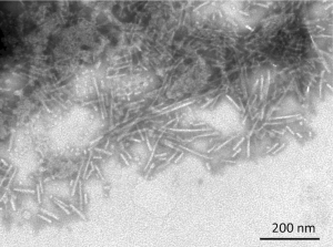
[Image from: StressMarq website] TEM characterization of Beta Synuclein Protein PFFs (catalog# SPR-457).
Summary
Seed amplification assays are effective tools for detecting fibrillar alpha synuclein aggregates in clinical samples and are capable of supporting the diagnosis of Parkinson’s disease. To improve upon the traditional seed amplification assay, researchers developed the quiescent seed amplification assay by eliminating the need for separate sonication or shaking steps, which are typically used to promote fibril amplification through cyclic fragmentation. By removing these mechanical steps, the QSAA reduces the risk of false positives and simplifies the procedure, requiring only a temperature-controlled fluorescence reader.
This enhanced assay offers a streamlined and sensitive approach for detecting pathological alpha synuclein, making it suitable for use with both brain and skin biopsy samples. The ability to analyze skin biopsies is particularly valuable, as it increases the accessibility of diagnostic testing by enabling less invasive sample collection.
Overall, the advancements introduced by the QSAA position it as a promising diagnostic tool for Parkinson’s disease. Its high sensitivity, specificity, and broader applicability could significantly improve early detection and clinical decision-making.
Related StressMarq products
StressMarq is committed to manufacturing high-quality, innovative reagents that support neurodegenerative disease research. These include a range of oligomeric, fibrillar and monomeric protein preparations, including alpha synuclein, beta synuclein, gamma synuclein, amyloid beta and tau. Visit our website for more information, including the latest scientific publications using our specialized protein constructs.
References
-
-
α-Synuclein seed amplification assays for diagnosing synucleinopathies: The way forward. Bellomo, G. et al. Neurology. 2022.
-
Ultrasensitive detection of aggregated α‑synuclein using quiescent seed amplification assay for the diagnosis of Parkinson’s disease. Mao, H. et al. Transl Neurodegener. 2025.
-
Leave a Reply