Discovery through Partnership | Excellence through Quality


Properties
| Storage Buffer | PBS pH 7.4, 50% glycerol, 0.09% Sodium azide *Storage buffer may change when conjugated |
| Storage Temperature | -20ºC, Conjugated antibodies should be stored according to the product label |
| Shipping Temperature | Blue Ice or 4ºC |
| Purification | Protein G Purified |
| Clonality | Monoclonal |
| Clone Number | 3C11 |
| Isotype | IgG1 |
| Specificity | Binds within the 81-130 aa region of human alpha synuclein, determined using WB and ELISA with corresponding peptides. Detects ~14 kDa band corresponding to Alpha synuclein. Band at ~30 kDa is a dimer. |
| Cite This Product | StressMarq Biosciences Cat# SMC-530, RRID: AB_2703053 |
| Certificate of Analysis | A 1:1000 dilution of SMC-530 was sufficient for detection of Alpha Synuclein in 15 µg of human brain cell lysate by ECL immunoblot analysis using goat anti-mouse IgG:HRP as the secondary antibody. |
Biological Description
| Alternative Names | Alpha Synuclein, α-Synuclein, SNCA, Snca, SYN, alphaSYN, NACP, Non-A beta component of AD amyloid, Non-A4 component of amyloid precursor, Parkinson disease familial 1, PARK1, PARK 1, PARK4, PARK 4, Parkinson disease (autosomal dominant, Lewy body) 4 |
| Research Areas | Alzheimer's Disease, Neurodegeneration, Neuroscience, Parkinson's Disease, Synuclein, Tangles & Tau, Multiple System Atrophy |
| Cellular Localization | Cell Junction, Cytoplasm, Cytosol, Membrane, Nucleus, Synapse |
| Accession Number | NP_000336.1 |
| Gene ID | 6622 |
| Swiss Prot | P37840 |
| Scientific Background | Alpha-synuclein (SNCA) is a neuronal protein predominantly expressed in the brain, where it is highly concentrated at presynaptic nerve terminals, suggesting a key role in synaptic transmission and plasticity (1). In addition to its synaptic localization, alpha-synuclein is abundantly expressed in mitochondria-rich brain regions, including the olfactory bulb, hippocampus, striatum, and thalamus, implicating it in mitochondrial function and energy metabolism (2). Functionally, alpha-synuclein has been shown to interact significantly with tubulin (3), indicating a potential role as a microtubule-associated protein involved in cytoskeletal organization and intracellular transport. This interaction may be critical for maintaining neuronal structure and function, particularly in long-range axonal projections. Importantly, alpha-synuclein is essential for normal cognitive development. Genetic or functional inactivation of SNCA has been associated with impairments in spatial learning and working memory, underscoring its role in higher-order brain functions (4). Given its physiological importance and pathological aggregation in disorders such as Parkinson’s disease and dementia with Lewy bodies, alpha-synuclein remains a focal point in neurodegenerative disease research. Understanding its normal function and mechanisms of misfolding is vital for developing targeted diagnostics and therapeutics. |
| References |
1. “Genetics Home Reference: SNCA”. US National Library of Medicine. (2013). 2. Zhang L., et al. (2008) Brain Res. 1244: 40-52. 3. Alim M.A., et al. (2002) J Biol Chem. 277(3): 2112-2117. 4. Kokhan V.S., Afanasyeva M.A., Van'kin G. (2012) Behav. Brain. Res. 231(1): 226-230. 5. Spillantini M.G., et al. (1997) Nature. 388(6645): 839-840. 6. Mezey E., et al. (1998) Nat Med. 4(7): 755-757. |
Product Images
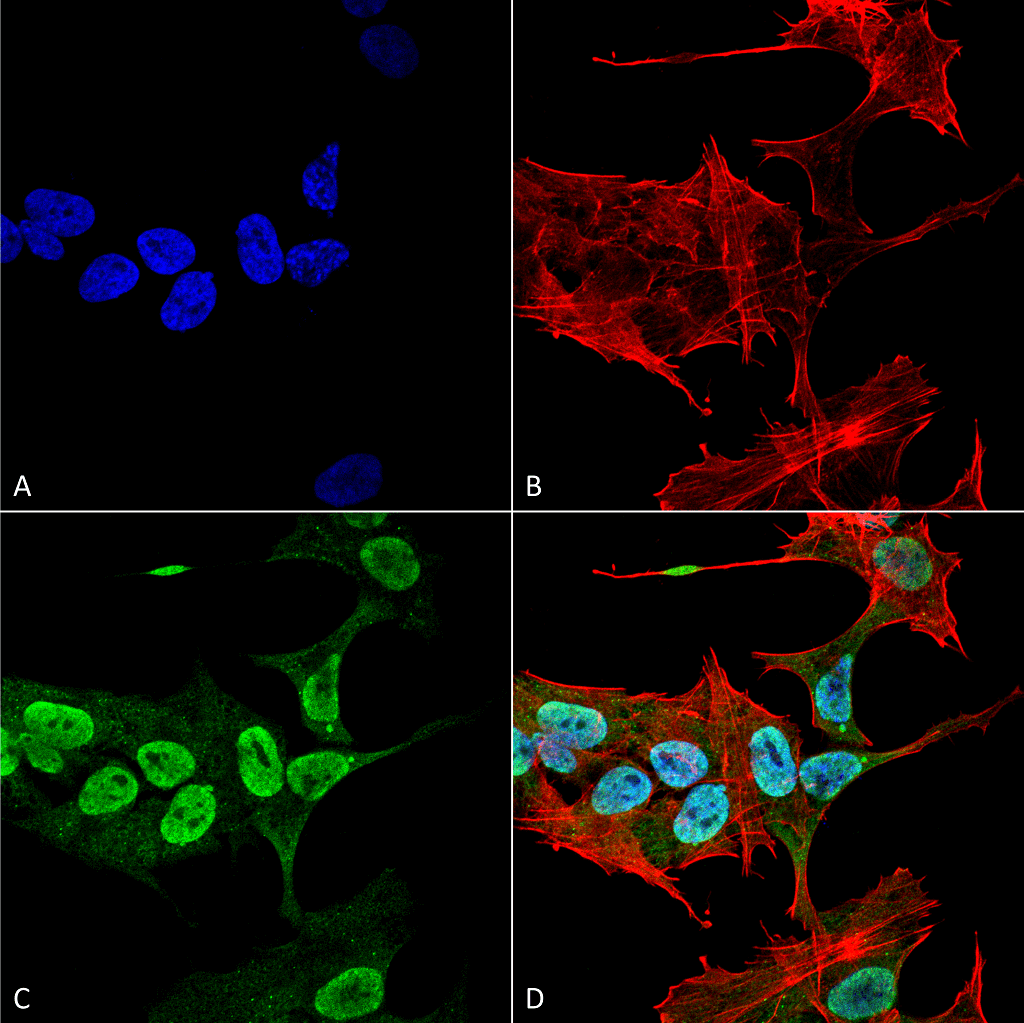
Immunocytochemistry/Immunofluorescence analysis using Mouse Anti-Alpha Synuclein Monoclonal Antibody, Clone 3C11 (SMC-530). Tissue: Neuroblastoma cell line (SK-N-BE). Species: Human. Fixation: 4% Formaldehyde for 15 min at RT. Primary Antibody: Mouse Anti-Alpha Synuclein Monoclonal Antibody (SMC-530) at 1:100 for 60 min at RT. Secondary Antibody: Goat Anti-Mouse ATTO 488 at 1:200 for 60 min at RT. Counterstain: Phalloidin Texas Red F-Actin stain; DAPI (blue) nuclear stain at 1:1000, 1:5000 for 60 min at RT, 5 min at RT. Localization: Cytoplasm: weak; Nucleus: Med. Magnification: 60X. (A) DAPI (blue) nuclear stain. (B) Phalloidin Texas Red F-Actin stain. (C) Alpha Synuclein Antibody. (D) Composite.
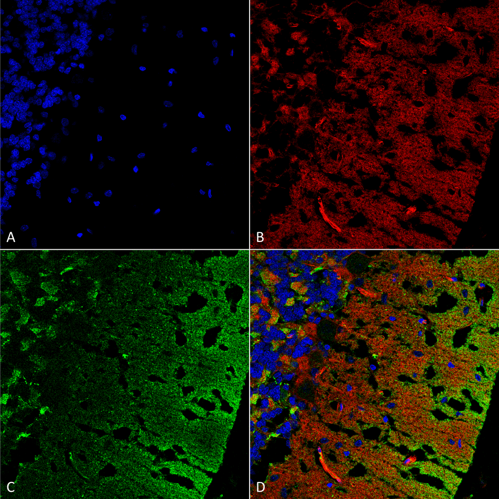
Immunohistochemistry analysis using Mouse Anti-Alpha Synuclein Monoclonal Antibody, Clone 3C11 (SMC-530). Tissue: cerebellum. Species: Rat. Fixation: Formalin fixed, paraffin embedded. Primary Antibody: Mouse Anti-Alpha Synuclein Monoclonal Antibody (SMC-530) at 1:25 for 1 hour at RT. Secondary Antibody: Goat Anti-Mouse IgG: Alexa Fluor 488. Counterstain: Actin-binding Phalloidin-Alexa Fluor 633; DAPI (blue) nuclear stain. Magnification: 63X. (A) DAPI (blue) nuclear stain. (B) Phalloidin Alexa Fluor 633 F-Actin stain. (C) Alpha Synuclein Antibody (D) Composite.
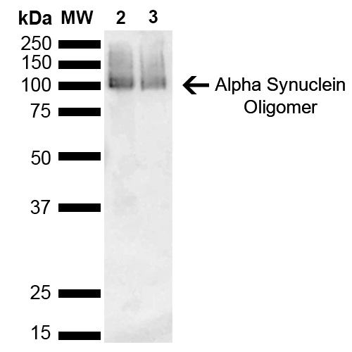
Western Blot analysis of Human Brain showing detection of 14 kDa Alpha Synuclein protein using Mouse Anti-Alpha Synuclein Monoclonal Antibody, Clone 3C11 (SMC-530). Lane 1: Molecular Weight Ladder (MW). Lane 2: Parkinson brain cell lystate. Lane 3: Human brain cell lysate. Load: 15 µg. Block: 5% Skim Milk in 1X TBST. Primary Antibody: Mouse Anti-Alpha Synuclein Monoclonal Antibody (SMC-530) at 1:1000 for 2 hours at RT. Secondary Antibody: Goat Anti-Mouse HRP:IgG at 1:3000 for 1 hour at RT. Color Development: ECL solution (Super Signal West Pico) for 5 min in RT. Predicted/Observed Size: 14 kDa. Other Band(s): 100 kDa (oligomer).
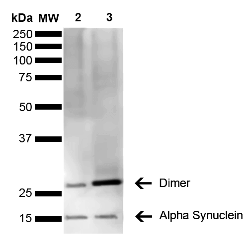
Western Blot analysis of Mouse, Rat Brain showing detection of 14 kDa Alpha Synuclein protein using Mouse Anti-Alpha Synuclein Monoclonal Antibody, Clone 3C11 (SMC-530). Lane 1: Molecular Weight Ladder (MW). Lane 2: Mouse brain cell lysate. Lane 3: Rat brain cell lysate. Load: 15 µg. Block: 5% Skim Milk in 1X TBST. Primary Antibody: Mouse Anti-Alpha Synuclein Monoclonal Antibody (SMC-530) at 1:1000 for 2 hours at RT. Secondary Antibody: Goat Anti-Mouse HRP:IgG at 1:3000 for 1 hour at RT. Color Development: ECL solution (Super Signal West Pico) for 5 min in RT. Predicted/Observed Size: 14 kDa. Other Band(s): ~30 kDa (dimer).
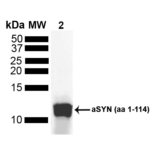
Western Blot analysis of Human Truncated Alpha Synuclein Protein showing detection of Alpha Synuclein protein using Mouse Anti-Alpha Synuclein Monoclonal Antibody, Clone 3C11 (SMC-530). Lane 1: MW Ladder. Lane 2: hASYN aa 1-114 (5 uL). Load: 5 uL. Block: 5% Skim Milk Powder in TBST. Primary Antibody: Mouse Anti-Alpha Synuclein Monoclonal Antibody (SMC-530) at 1:1000 for 2 hours at RT with shaking . Secondary Antibody: Goat anti-mouse IgG:HRP at 1:4000 for 1 hour at RT with shaking . Color Development: Chemiluminescent for HRP (Moss) for 5 min in RT.
 Powered by Bioz
StressMarq Biosciences :
Based on validation through cited publications.