Discovery through Partnership | Excellence through Quality


Properties
| Storage Buffer | PBS pH 7.4, 50% glycerol, 0.09% Sodium azide *Storage buffer may change when conjugated |
| Storage Temperature | -20ºC, Conjugated antibodies should be stored according to the product label |
| Shipping Temperature | Blue Ice or 4ºC |
| Purification | Protein G Purified |
| Clonality | Monoclonal |
| Clone Number | 1B8 |
| Isotype | IgG2a |
| Specificity | Detects ~33 kDa protein. |
| Cite This Product | StressMarq Biosciences Cat# SMC-267, RRID: AB_2699952 |
| Certificate of Analysis | A 1:1000 dilution of SMC-267 was sufficient for detection of CD74 in 15 µg of B-Cell Lymphoma cell line Raji by ECL immunoblot analysis using Goat Anti-Mouse IgG:HRP as the secondary Antibody. |
Biological Description
| Alternative Names | CD74, DHLAG, HLA DR gamma, HLADG, p33, p35, Protein 41, CD74 antigen (invariant polypeptide of major histocompatibility complex, class II antigen-associated), CD74 antigen, CD74 molecule, CD74 molecule, major histocompatibility complex, class II invariant chain, CLIP, Gamma chain of class II antigens, HG2A_HUMAN, HLA class II histocompatibility antigen gamma chain, HLA DR antigens associated invariant chain, HLA-DR antigens-associated invariant chain, HLA-DR-gamma, HLADR antigens associated invariant chain, Ia antigen associated invariant chain, Ia antigen-associated invariant chain, Ia associated invariant chain, Ia gamma, Ii, Invariant polypeptide of major histocompatibility complex class II antigen associated, la-gamma, Major histocompatibility complex class II invariant chain, MHC HLA DR gamma chain, MHC HLA-DR gamma chain |
| Research Areas | Adaptive Immunity, B Cell CD Markers, B Cells, Immunology |
| Cellular Localization | Cell membrane, Endoplasmic Reticulum, Endoplasmic reticulum membrane, Endosome, Golgi apparatus, Lysosome |
| Accession Number | NP_001020329.1 |
| Gene ID | 972 |
| Swiss Prot | P04233 |
| Scientific Background |
CD74, also known as the invariant chain, is a non-polymorphic type II transmembrane protein primarily recognized for its role as a chaperone for MHC class II molecules. It facilitates the proper folding, ER exit, and trafficking of MHC class II complexes to endosomal compartments, where it also contributes to peptide editing and antigen presentation. While traditionally associated with professional antigen-presenting cells, CD74 expression extends to non-immune cells under inflammatory conditions, including epithelial and neural cells. This broader expression pattern has important implications for neuroinflammation and neurodegenerative disease. Beyond its classical role, CD74 also functions as a high-affinity receptor for macrophage migration inhibitory factor (MIF), a pro-inflammatory cytokine implicated in neurodegenerative disorders such as Alzheimer’s disease and multiple sclerosis. CD74-MIF signaling activates downstream pathways involved in cell survival, immune activation, and neuroinflammation—key processes in the progression of neurodegeneration. Notably, CD74 has a very short surface half-life (under 10 minutes), rapidly internalizing upon ligand binding. This property, combined with its restricted expression in healthy tissues, makes CD74 an attractive target for therapeutic intervention in both cancer and neuroinflammatory conditions. As a molecule at the intersection of immune regulation and neuronal health, CD74 is gaining recognition as a critical player in the pathophysiology of neurodegenerative diseases. |
| References |
1. Becker-Hermann, S., Arie, G., Medvedovsky H, Kerem A, and Shachar I. (2005) Mol Bio Cell. 16(11):5061-9. 2. Barrera CA., et al (2005) J Histochem Cytochem 53 (12): 1481-9. 3. Starlets D., et al. (2006) Blood. 107 (12): 4807-4816. 4. Burton JD., et al. (2004). Clin Cancer Res. 10(19): 6606-11. 5. Denzin L.K., Hammond, C. and Cresswell, P. (1996) J. Exp. Med. 184: 2153-2165. 6. Denzin L.K., Robbins N.F., Carboy-Newcomb C. and Cresswell P. (1994) Immunity 1: 595-606. |
Product Images
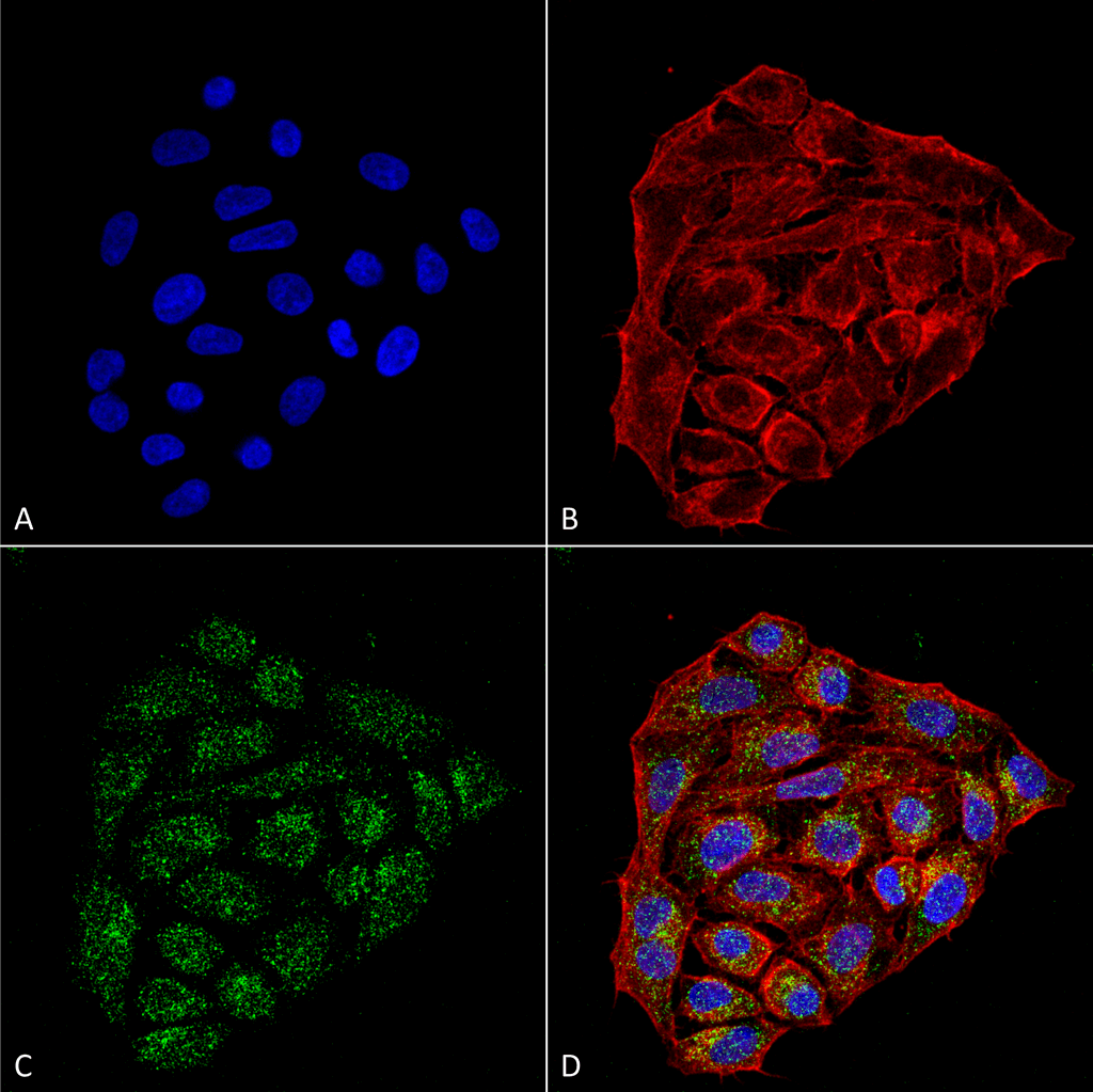
Immunocytochemistry/Immunofluorescence analysis using Mouse Anti-CD74 Monoclonal Antibody, Clone 1B8 (SMC-267). Tissue: Cervical cancer cell line (HeLa). Species: Human. Fixation: 4% Formaldehyde for 15 min at RT. Primary Antibody: Mouse Anti-CD74 Monoclonal Antibody (SMC-267) at 1:100 for 60 min at RT. Secondary Antibody: Goat Anti-Mouse ATTO 488 at 1:200 for 60 min at RT. Counterstain: Phalloidin Texas Red F-Actin stain; DAPI (blue) nuclear stain at 1:1000, 1:5000 for 60 min at RT, 5 min at RT. Localization: Cell membrane, Endoplasmic Reticulum, Golgi apparatus. Magnification: 60X. (A) DAPI (blue) nuclear stain. (B) Phalloidin Texas Red F-Actin stain. (C) CD74 Antibody. (D) Composite.
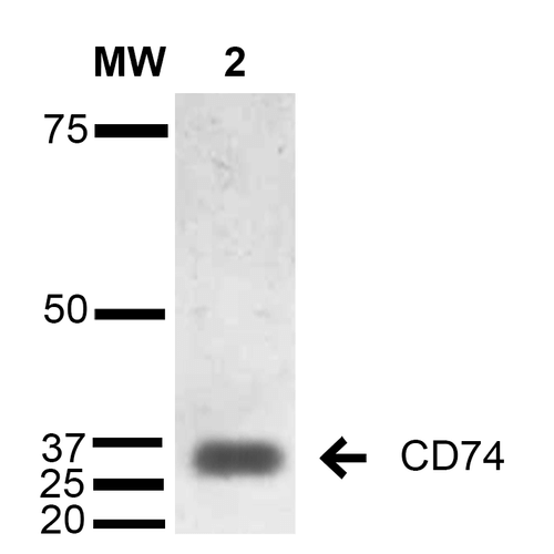
Western Blot analysis of Human Lymphoblastoid cell line (Raji) showing detection of 33-35 kDa CD74 protein using Mouse Anti-CD74 Monoclonal Antibody, Clone 1B8 (SMC-267). Lane 1: Molecular Weight Ladder (MW). Lane 2: Raji cell lysate. Load: 15 µg. Block: 5% Skim Milk in TBST. Primary Antibody: Mouse Anti-CD74 Monoclonal Antibody (SMC-267) at 1:1000 for 2 hours at RT. Secondary Antibody: Goat Anti-Mouse IgG: HRP at 1:1000 for 60 min at RT. Color Development: ECL solution for 5 min in RT. Predicted/Observed Size: 33-35 kDa.
 Powered by Bioz
Reviews
There are no reviews yet.