Discovery through Partnership | Excellence through Quality


Properties
| Storage Buffer | PBS pH7.4, 50% glycerol, 0.09% sodium azide *Storage buffer may change when conjugated |
| Storage Temperature | -20ºC, Conjugated antibodies should be stored according to the product label |
| Shipping Temperature | Blue Ice or 4ºC |
| Purification | Protein G Purified |
| Clonality | Monoclonal |
| Clone Number | 1F2-H5 |
| Isotype | IgG2a Kappa |
| Specificity | Detects ~73kDa. Does not cross react with HSP70. |
| Cite This Product | StressMarq Biosciences Cat# SMC-151, RRID: AB_2120165 |
| Certificate of Analysis | 1 µg/ml of SMC-151 was sufficient for detection of HSC70 in 10 µg of HeLa lysate by colorimetric immunoblot analysis using Goat anti-mouse IgG:HRP as the secondary antibody. |
Biological Description
| Alternative Names | HSPA8, HSC70, HSP73, Heat shock cognate 71 kDa protein, Heat shock 70 kDa protein 8, HSP71, HSC71, HSC54, HSC73, HSPA10, LAP1, NIP71 |
| Research Areas | Cancer, Cell Signaling, Chaperone Proteins, Heat Shock, Protein Trafficking |
| Cellular Localization | Cytoplasm, Melanosome |
| Accession Number | NP_006588.1 |
| Gene ID | 3312 |
| Swiss Prot | P11142 |
| Scientific Background | The HSP70 family comprises highly conserved, heat-inducible 70-kDa proteins that play a pivotal role in maintaining cellular proteostasis, particularly under stress conditions. Encoded by a multigene family in most eukaryotes, HSP70 proteins are ubiquitously distributed across cellular compartments—including the cytosol, nucleus, mitochondria, endoplasmic reticulum, and chloroplasts—and are also present in prokaryotes. Their evolutionary conservation, with over 50% sequence identity, underscores their essential biological functions. HSP70 proteins exhibit high-affinity ATP binding and possess ATPase activity, which is enhanced upon interaction with unfolded proteins and synthetic peptides. Structurally, the N-terminal domain mediates ATP binding, while the C-terminal domain governs substrate interaction. This dual-domain architecture enables HSP70s to act as molecular chaperones, facilitating protein folding, preventing aggregation, and promoting recovery from proteotoxic stress. In the context of neurodegenerative diseases, inducible HSP70 (HSP72) and its constitutively expressed counterpart HSC70 are of particular interest. Following cellular stress, such as heat shock, HSP72 is rapidly upregulated and forms ATP-dependent complexes with HSC70. This interaction is critical for restoring centrosomal integrity and function—processes often disrupted in neurodegenerative pathologies like Alzheimer’s and Parkinson’s disease. Given their central role in protein quality control and cellular resilience, HSP70 proteins are emerging as promising therapeutic targets in neuroscience and neurodegeneration research. |
| References |
1. Brown C.L. et al. (1993) J.Cell Biol., 120 (5): 1101-1112. 2. Boorstein W.R., Ziegelhoffer T., and Craig E.A. (1993)J. Mol. Evol. 38(1): 1-17. 3. Rothman J. (1989), Cell 59: 591-601. 4. DeLuca-Flaherty et al. (1990) Cell 62: 875-887. 5. Bork P., Sander C., and Valencia A. (1992) Proc. Nut1Acad. Sci. USA 89: 7290-7294. 6. Brown C.L. et al. (1996) J. Biol. Chem. 271(2): 833-840. |
Product Images
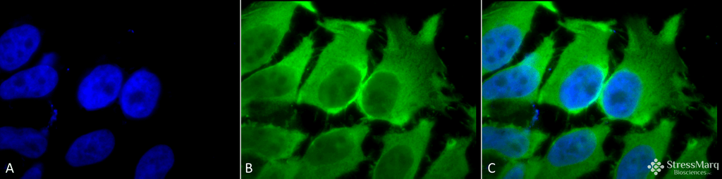
Immunocytochemistry/Immunofluorescence analysis using Mouse Anti-Hsc70 (Hsp73) Monoclonal Antibody, Clone 1F2-H5 (SMC-151). Tissue: Heat Shocked cervical cancer cells (HeLa). Species: Human. Fixation: 2% Formaldehyde for 20 min at RT. Primary Antibody: Mouse Anti-Hsc70 (Hsp73) Monoclonal Antibody (SMC-151) at 1:100 for 12 hours at 4°C. Secondary Antibody: FITC Goat Anti-Mouse (green) at 1:200 for 2 hours at RT. Counterstain: DAPI (blue) nuclear stain at 1:40000 for 2 hours at RT. Localization: Cytoplasm. Melanosome. Localizes to nucleus upon heat shock. Magnification: 100x. (A) DAPI (blue) nuclear stain. (B) Anti-Hsc70 (Hsp73) Antibody. (C) Composite. Heat Shocked at 42°C for 1h.
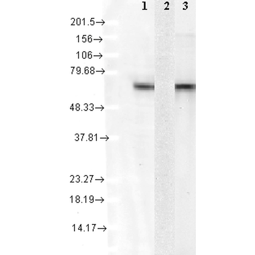
Western Blot analysis of Human Cell lysates showing detection of Hsc70 protein using Mouse Anti-Hsc70 Monoclonal Antibody, Clone 1F2-H5 (SMC-151). Load: 15 µg. Block: 1.5% BSA for 30 minutes at RT. Primary Antibody: Mouse Anti-Hsc70 Monoclonal Antibody (SMC-151) at 1:1000 for 2 hours at RT. Secondary Antibody: Sheep Anti-Mouse IgG: HRP for 1 hour at RT. 1: mix of 10 different human cell lines, 2: Hsp72 recombinant protein, and 3: Hsc70(Hsp73) recombinant protein.
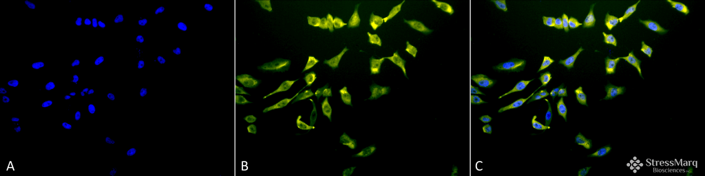
Immunocytochemistry/Immunofluorescence analysis using Mouse Anti-Hsc70 (Hsp73) Monoclonal Antibody, Clone 1F2-H5 (SMC-151). Tissue: Heat Shocked cervical cancer cells (HeLa). Species: Human. Fixation: 2% Formaldehyde for 20 min at RT. Primary Antibody: Mouse Anti-Hsc70 (Hsp73) Monoclonal Antibody (SMC-151) at 1:100 for 12 hours at 4°C. Secondary Antibody: R-PE Goat Anti-Mouse (yellow) at 1:200 for 2 hours at RT. Counterstain: DAPI (blue) nuclear stain at 1:40000 for 2 hours at RT. Localization: Cytoplasm. Melanosome. Localizes to nucleus upon heat shock. Magnification: 20x. (A) DAPI (blue) nuclear stain. (B) Anti-Hsc70 (Hsp73) Antibody. (C) Composite. Heat Shocked at 42°C for 1h.
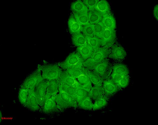
Immunocytochemistry/Immunofluorescence analysis using Mouse Anti-Hsc70 Monoclonal Antibody, Clone 1F2-H5 (SMC-151). Tissue: HaCaT cells. Species: Human. Fixation: Cold 100% methanol for 10 minutes at -20°C. Primary Antibody: Mouse Anti-Hsc70 Monoclonal Antibody (SMC-151) at 1:100 for 1 hour at RT. Secondary Antibody: FITC Goat Anti-Mouse (green) at 1:50 for 1 hour at RT. Localization: Bright cytoplasmic staining, duller nuclear staining.
 Powered by Bioz
StressMarq Biosciences :
Based on validation through cited publications.