Discovery through Partnership | Excellence through Quality


Properties
| Storage Buffer | PBS, 50% glycerol, 0.09% sodium azide *Storage buffer may change when conjugated |
| Storage Temperature | -20ºC, Conjugated antibodies should be stored according to the product label |
| Shipping Temperature | Blue Ice or 4ºC |
| Purification | Protein G Purified |
| Clonality | Monoclonal |
| Clone Number | 39B6 |
| Isotype | IgG2a |
| Specificity | Recognizes 3-nitrotyrosine moieties. No detectable cross-reactivity with non-nitrated tyrosine. Not species specific. |
| Cite This Product | StressMarq Biosciences Cat# SMC-154, RRID: AB_904533 |
| Certificate of Analysis | 0.7 µg/ml of SMC-154 was sufficient for detection of 5 µg SIN-1 treated BSA by Western Blot analysis using Goat anti-mouse IgG:HRP as the secondary antibody. |
Biological Description
| Alternative Names | Nitrotyrosine, Nitro tyrosine, 3-Nitrotyrosine |
| Research Areas | Alzheimer's Disease, Cancer, Cell Signaling, Neurodegeneration, Neuroscience, Nitration, Oxidative Stress, Parkinson's Disease, Post-translational Modifications |
| Scientific Background | Nitrotyrosine is a post-translational modification formed by the nitration of tyrosine residues, often mediated by reactive nitrogen species such as peroxynitrite. It serves as a biomarker of nitrosative stress and is increasingly recognized for its role in neurodegenerative disease pathology. Nitrotyrosine-modified proteins are frequently detected in Alzheimer’s disease, Parkinson’s disease, and ALS, where they correlate with inflammation-induced tissue injury. Enzymes such as myeloperoxidase, cytochrome P450s, and superoxide dismutase catalyze tyrosine nitration, disrupting protein function and contributing to neuronal damage. As a marker of oxidative and nitrosative stress, nitrotyrosine provides mechanistic insight into redox imbalance and may serve as a diagnostic and therapeutic target in neurodegeneration. |
| References |
1. Girault I. et al. (2001). Free Radical Biology and Medicine, 31 (11): 1375-1387. 2. Gow AJ, Farkouh CR, Munson DA, Posencheq MA, and Ischiropoulos H. (2004). Am J Physiol Lung Cell Mol Physiol. 287(2): L262-8. 3. Takemoto K. et al (2007). Acta Med Okayama 61(1): 17-30. 4. Reynolds MR. et al. (2006) J Nerosci. 26(42): 10636-45. 5. Pfister H., et al. (2002) Vet Pathol. 39: 190-199. 6. Khan J. et al. (1998) Biochem J. 330(2): 795-801. |
Product Images
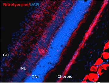
Immunohistochemistry analysis using Mouse Anti-Nitrotyrosine Monoclonal Antibody, Clone 39B6 (SMC-154). Tissue: Retinal Injury Model. Species: Mouse. Primary Antibody: Mouse Anti-Nitrotyrosine Monoclonal Antibody (SMC-154) at 1:1000. Secondary Antibody: Alexa Fluor 594 Goat Anti-Mouse (red). Courtesy of: Dr. Rajashekhar Gangaraju, University of Indiana, Department of Ophthalmology, Eugene and Marilyn Glick Eye Institute.
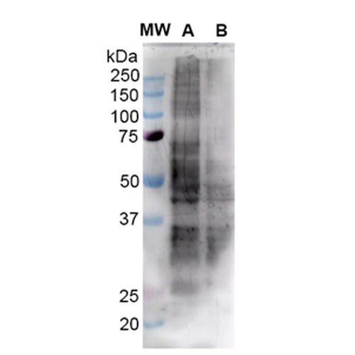
Western Blot analysis of Human HEK293 cells showing detection of Nitrotyrosine protein using Mouse Anti-Nitrotyrosine Monoclonal Antibody, Clone 39B6 (SMC-154). Lane 1: MW Ladder. Lane A: Nitrosylated-HEK293 (15uL). Lane B: HEK293 (15 ug). Block: 5% Skim Milk Powder in TBST. Primary Antibody: Mouse Anti-Nitrotyrosine Monoclonal Antibody (SMC-154) diluted in 1.5% BSA and TBST for 1 hours at RT with shaking . Secondary Antibody: Goat anti-mouse IgG: HRP at 1:4000 for 1 hour at RT with shaking . Predicted/Observed Size: Multiple Bands.
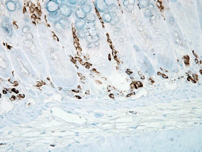
Immunohistochemistry analysis using Mouse Anti-Nitrotyrosine Monoclonal Antibody, Clone 39B6 (SMC-154). Tissue: inflamed colon. Species: Mouse. Fixation: Formalin. Primary Antibody: Mouse Anti-Nitrotyrosine Monoclonal Antibody (SMC-154) at 1:1000000 for 12 hours at 4°C. Secondary Antibody: Biotin Goat Anti-Mouse at 1:2000 for 1 hour at RT. Counterstain: Mayer Hematoxylin (purple/blue) nuclear stain at 200 µl for 2 minutes at RT. Magnification: 40x. With anti-microbial. This image was produced using an amplifying IHC wash buffer. The antibody has therefore been diluted more than is recommended for other applications.
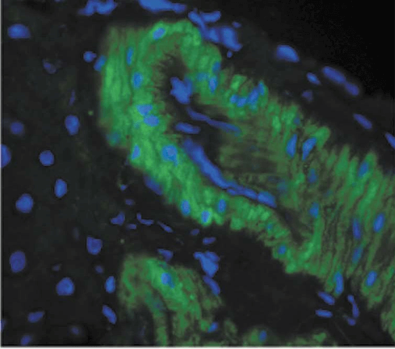
Immunohistochemistry analysis using Mouse Anti-Nitrotyrosine Monoclonal Antibody, Clone 39B6 (SMC-154). Tissue: liver tissue . Species: Rat. Primary Antibody: Mouse Anti-Nitrotyrosine Monoclonal Antibody (SMC-154) at 1:1000. Secondary Antibody: FITC Goat Anti-Mouse (green).
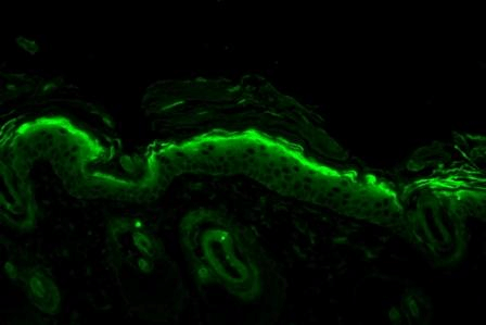
Immunohistochemistry analysis using Mouse Anti-Nitrotyrosine Monoclonal Antibody, Clone 39B6 (SMC-154). Tissue: backskin. Species: Mouse. Fixation: Bouin’s Fixative and paraffin-embedded. Primary Antibody: Mouse Anti-Nitrotyrosine Monoclonal Antibody (SMC-154) at 1:100 for 1 hour at RT. Secondary Antibody: FITC Goat Anti-Mouse (green) at 1:50 for 1 hour at RT. Backskin obtained from transgenic mice.
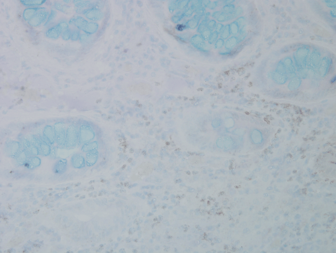
Immunohistochemistry analysis using Mouse Anti-Nitrotyrosine Monoclonal Antibody, Clone 39B6 (SMC-154). Tissue: colon carcinoma. Species: Human. Fixation: Formalin. Primary Antibody: Mouse Anti-Nitrotyrosine Monoclonal Antibody (SMC-154) at 1:25000 for 12 hours at 4°C. Secondary Antibody: Biotin Goat Anti-Mouse at 1:2000 for 1 hour at RT. Counterstain: Mayer Hematoxylin (purple/blue) nuclear stain at 200 µl for 2 minutes at RT. Magnification: 40x.
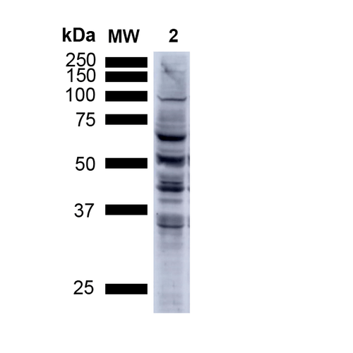
Western Blot analysis of Human A549 cells showing detection of Multiple Bands Nitrotyrosine protein using Mouse Anti-Nitrotyrosine Monoclonal Antibody, Clone 39B6 (SMC-154). Lane 1: MW ladder. Lane 2: Human A549 Cells 15 ug). Load: 15 ug. Block: 5% Skim Milk Powder in TBST. Primary Antibody: Mouse Anti-Nitrotyrosine Monoclonal Antibody (SMC-154) at 1:1000 for 2.5 hours at RT with shaking . Secondary Antibody: Goat anti-mouse IgG:HRP at 1:1000 for 1 hour at RT with shaking . Color Development: Chemiluminescent for HRP (Moss) for 5 min in RT. Predicted/Observed Size: Multiple Bands.
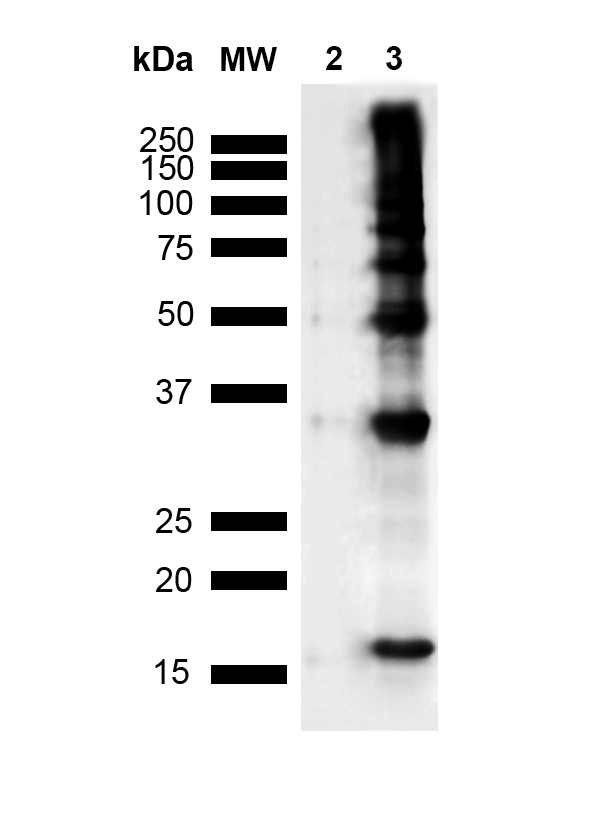
Western Blot analysis of Human Recombinant Protein showing detection of Multiple Bands Nitrotyrosine protein using Mouse Anti-Nitrotyrosine Monoclonal Antibody, Clone 39B6 (SMC-154). Lane 1: MW Ladder. Lane 2: hASYN Monomer (3.84 ug). Lane 3: Nitrosylated hASYN (3.84 ug).. Block: 5% Skim Milk Powder in TBST. Primary Antibody: Mouse Anti-Nitrotyrosine Monoclonal Antibody (SMC-154) at 1:1000 for 2 hours at RT with shaking . Secondary Antibody: Goat anti-mouse IgG:HRP at 1:4000 for 2 hour at RT with shaking . Color Development: Chemiluminescent for HRP (Moss) for 5 min in RT. Predicted/Observed Size: Multiple Bands.
 Powered by Bioz
StressMarq Biosciences :
Based on validation through cited publications.